[IF=7.561] Ma Y. et al. Photobiomodulation Attenuates Neurotoxic Polarization of Macrophages by Inhibiting the Notch1-HIF-1α/NF-κB Signalling Pathway in Mice With Spinal Cord Injury.. Front Immunol. 2022 Mar;13:816952-816952 WB ; Mouse.
[IF=3.575] Chang Hao. et al. Hypoxic preconditioning improves the survival and pro-angiogenic capacity of transplanted human umbilical cord mesenchymal stem cells via HIF-1α signaling in a rat model of bronchopulmonary dysplasia. Biochem Bioph Res Co. 2022 Mar;: WB ; Rat.
[IF=0.5] Talwar, Harvinder, et al. ""The dataset describes: HIF-1 α expression and LPS mediated cytokine production in MKP-1 deficient bone marrow derived murine macrophages. Data in Brief (2017). 14:56-61.." WB ; Mouse.
[IF=5.595] Liu T et al. MicroRNA-493 targets STMN-1 and promotes hypoxia-induced epithelial cell cycle arrest in G/M and renal fibrosis. (2018) FASEB J. Sep 05 WB ; Mouse.
[IF=2.766] Dao DT et al. A paradoxical method to enhance compensatory lung growth: Utilizing a VEGF inhibitor.(2018) PLoS One. WB ; Mouse.
[IF=4.556] Yang X et al. Synthesis and bioevaluation of novel 18FDG-conjugated 2-nitroimidazole derivatives for tumor-hypoxia imaging. Mol Pharm. 2019 May 6;16(5):2118-2128. IHC&IF ; Mouse&Human.
[IF=2.72] Jiebin Li. et al. Targeted Temperature Management Suppresses Hypoxia-Inducible Factor-1α and Vascular Endothelial Growth Factor Expression in a Pig Model of Cardiac Arrest. 2021 Jan 05 WB ; Pig.
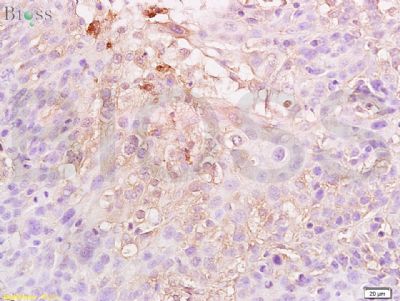
[IF=6.843] Zhuowen Yang. et al. Ferrite-encapsulated nanoparticles with stable photothermal performance for multimodal imaging-guided atherosclerotic plaque neovascularization therapy. Biomater Sci-Uk. 2021 Jul;: IHC ; Rabbit.
[IF=3.738] Zong, Jinxin. et al. Lithium Chloride Promotes Milk Protein and Fat Synthesis in Bovine Mammary Epithelial Cells via HIF-1α and β-Catenin Signaling Pathways. Biol Trace Elem Res. 2022 Jan;:1-16 WB ; Bovine.
[IF=14.65] Liu, Hao-Yu. et al. Distinct B cell subsets in Peyer’s patches convey probiotic effects by Limosilactobacillus reuteri. Microbiome. 2021 Dec;9(1):1-18 FC ; Mouse.
[IF=2.447] Xuejun Dai. et al. Comparison of the differentiation abilities of bone marrow‑derived mesenchymal stem cells and adipose‑derived mesenchymal stem cells toward nucleus pulposus‑like cells in three‑dimensional culture. Exp Ther Med. 2021 Sep;22(3):1-9 WB ; Rat.
[IF=5.923] Ayaka Ito. et al. Elevation of Chemosensitivity of Lung Adenocarcinoma A549 Spheroid Cells by Claudin-2 Knockdown through Activation of Glucose Transport and Inhibition of Nrf2 Signal. Int J Mol Sci. 2021 Jan;22(12):6582 WB ; Human.
[IF=10.652] Zhuo Liang. et al. Exosome derived from mesenchymal stem cells mediates hypoxia-specific BMP2 gene delivery and enhances bone regeneration. Chem Eng J. 2021 Oct;422:130084 IF ; Rabbit.
[IF=6.183] Renjie Luo. et al. An Albumin-binding Dimeric Prodrug Nanoparticle with Long Blood Circulation and Light-triggered Drug Release for Chemo-photodynamic Combination Therapy against Hypoxia-induced Metastasis of Lung Cancer. 2021 Mar 26 IHC ; Mouse.
[IF=6.895] Fangmei Zhang. et al. X-ray-triggered NO-released Bi–SNO nanoparticles: all-in-one nano-radiosensitizer with photothermal/gas therapy for enhanced radiotherapy. 2020 Aug 31 IF ; Human.
[IF=3.297] Yongjun Gao. et al. Study on the differential proteomics of rat hippocampal mitochondria during deep hypothermic circulatory arrest. Ann Transl Med. 2021 Feb; 9(4): 346 IHC ; Rat.
[IF=5.88] Rui X et al. Imperative and effective reversion of synovial hyperplasia and cartilage destruction in rheumatoid arthritis through multiple synergistic effects of O2 and Ca2+. Mater Sci Eng C Mater Biol Appl.2020 Sep;114:111058. IHC ; Rat.
[IF=3.361] Ouyang H et al. MicroRNA-208-5p regulates myocardial injury of sepsis mice via targeting SOCS2-mediated NF-κB/HIF-1α pathway. Int Immunopharmacol. 2020 Feb 18;81:106204. WB ; Mouse.
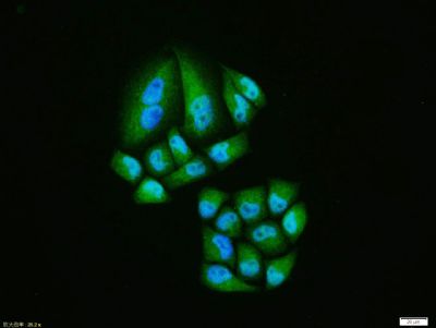
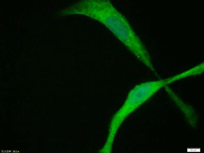
[IF=5.656] Duda P et al. The Reverse Warburg Effect is Associated with Fbp2-Dependent Hif1α Regulation in Cancer Cells Stimulated by Fibroblasts. Cells. 2020 Jan 14;9(1). pii: E205. ICF&Co-IP&WB ; Mouse&Human.
[IF=2.6] Wu TH et al. The Combination of Astragalus membranaceus and Angelica sinensis Inhibits Lung Cancer and Cachexia through Its Immunomodulatory Function. J Oncol. 2019 Nov 3;2019:9206951. WB ; Mouse.
[IF=3.414] Li ZH et al. You-Gui-Yin improved the reproductive dysfunction of male rats with chronic kidney disease via regulating the HIF1α-STAT5 pathway. J Ethnopharmacol. 2020 Jan 10;246:11248. WB ; Rat.
[IF=3.288] Chai D et al. β2-microglobulin has a different regulatory molecular mechanism between ER+ and ER- breast cancer with HER2.BMC Cancer. 2019 Mar 12;19(1):223. IHSLCP ; Human.
[IF=4.539] Talwar H et al. MEK2 Negatively Regulates Lipopolysaccharide-Mediated IL-1β Production through HIF-1α Expression. J Immunol. 2019 Mar 15;202(6):1815-1825. WB&IP ; Mouse.
[IF=1.41] Song et al. Effects of HSYA on the proliferation and apoptosis of MSCs exposed to hypoxic and serum deprivation conditions. (2018) Exp.Ther.Med. 15:5251-5260 WB ; Rat.
[IF=3.53] Woolf, Eric C., et al. "The Ketogenic Diet Alters the Hypoxic Response and Affects Expression of Proteins Associated with Angiogenesis, Invasive Potential and Vascular Permeability in a Mouse Glioma Model." PLOS ONE10.6 (2015): e0130357. WB ; Mouse.
[IF=3.869] Chen Guanyin. et al. Hypoxia-Induced Mesenchymal Stem Cells Exhibit Stronger Tenogenic Differentiation Capacities and Promote Patellar Tendon Repair in Rabbits. Stem Cells Int. 2020;2020:8822609 IF,WB ; Rabbit.
[IF=3] Meng-Meng ZHANG. et al. Protective effect of Pai-Nong-San against AOM/DSS-induced CAC in mice through inhibiting the Wnt signaling pathway. Chin J Nat Medicines. 2021 Dec;19:912 IHC ; Mouse.
[IF=8.071] Ze Zhang. et al. Oxygen sensors mediated HIF-1α accumulation and translocation: A pivotal mechanism of fine particles-exacerbated myocardial hypoxia injury. Environ Pollut. 2022 May;300:118937 WB,IF,IHC,IP ; Mouse,Rat.
[IF=6.244] Sun Q. et al. PKM2 Is the Target of a Multi-Herb-Combined Decoction During the Inhibition of Gastric Cancer Progression.. Front Oncol. 2021 Dec;11:767116-767116 WB ; Human.
[IF=4.375] Sun Tianyu. et al. The Human Positive Cofactor 4 is a Promising Chemotherapeutic Target in Lung Adenocarcinoma. J Oncol. 2021;2021:9958483 WB ; Human.
[IF=16.806] Jianting Yao. et al. Low-Intensity Focused Ultrasound-Responsive Ferrite-Encapsulated Nanoparticles for Atherosclerotic Plaque Neovascularization Theranostics. 2021 Aug 11 IF ; Rabbit.
[IF=8.128] Huaying Hou. et al. NIR-driven intracellular photocatalytic oxygen-supply on metallic molybdenum carbide@N-carbon for hypoxic tumor therapy. J Colloid Interf Sci. 2022 Feb;607:1 IHC ; Mouse.
[IF=5.037] Aslı Okan. et al. Immunoreactive definition of TNF- α, HIF-1 α, Kir6.2, Kir3.1 and M2 muscarinic receptor for cardiac and pancreatic tissues in a mouse model for type 1 diabetes. Life Sci. 2021 Aug;:119886 IHC ; Mouse.
[IF=4.451] Shiying Huang. et al. Jujube polysaccharides mitigated anemia in rats with chronic kidney disease: Regulation of short chain fatty acids release and erythropoietin production. J Funct Foods. 2021 Nov;86:104673 IHC ; Rat.
[IF=1.95] Xiaohua Du. et al. Expression and distribution of neuroglobin and hypoxia-inducible factor-1α in the adult yak telencephalon. 2021 Jun 19 WB ; Yak.
[IF=11.508] Rui Shao. et al. H3K36 methyltransferase NSD1 regulates chondrocyte differentiation for skeletal development and fracture repair. Bone Res. 2021 Jun;9(1):1-11 WB ; Mouse.
[IF=5.344] Ying Long. et al. A hybrid membrane coating nanodrug system against gastric cancer via VEGFR2/STAT3 signaling pathway. 2021 Apr 07 WB ; Human.
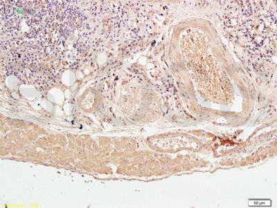
[IF=14.588] Donglin Xia. et al. Au–Hemoglobin Loaded Platelet Alleviating Tumor Hypoxia and Enhancing the Radiotherapy Effect with Low-Dose X-ray. Acs Nano. 2020;14(11):15654–15668 IHC ; Mouse.
[IF=2.842] Guangrui Chai. et al. NLRP3 Blockade Suppresses Pro-Inflammatory and Pro-Angiogenic Cytokine Secretion in Diabetic Retinopathy. Diabet Metab Synd Ob. 2020; 13: 3047–3058 WB ; Human.
[IF=4.105] Jacopo Di Gregorio. et al. UBXN7 cofactor of CRL3KEAP1 and CRL2VHL ubiquitin ligase complexes mediates reciprocal regulation of NRF2 and HIF-1α proteins. Bba-Mol Cell Res. 2021 Apr;1868:118963 WB ; Human.
[IF=10.317] Qi Sun. et al. Phototherapy and anti-GITR antibody-based therapy synergistically reinvigorate immunogenic cell death and reject established cancers. Biomaterials. 2021 Feb;269:120648 IHC ; Mouse.
[IF=2.91] Shou, Zhu, et al. "Expression and prognosis of FOXO3a and HIF-1?? in nasopharyngeal carcinoma."Journal of cancer research and clinical oncology 138.4 (2012): 585-593.. WB ; Human.
[IF=3.332] Sun Y et al. The function of Piezo1 in colon cancer metastasis and its potential regulatory mechanism. J Cancer Res Clin Oncol. 2020 May;146(5):1139-1152. IHC,WB ; human.
[IF=5.646] Shen Y et al. Tumor vasculature remolding by thalidomide increases delivery and efficacy of cisplatin. J Exp Clin Cancer Res. 2019 Oct 28;38(1):427. IHF-P ; Mouse.
[IF=3.743] Wang D et al. Effects of hypoxia and ASIC3 on nucleus pulposus cells: From cell behavior to molecularmechanism. Biomed Pharmacother. 2019 Jun 12;117:109061. WB ; Rabbit.
[IF=2.566] Yang Z et al. Tenascin-C is involved in promotion of cancer stemness via the Akt/HIF1ɑ axis in esophageal squamous cell carcinoma.Exp Mol Pathol. 2019 Mar 20. WB ; Human.
[IF=2.784] Yang D et al. Normobaric oxygen inhibits AQP4 and NHE1 expression in experimental focal ischemic stroke. (2018) Int. J. Mol. Med. WB ; Rat.
[IF=2.989] Ju X et al. Catalpol Promotes the Survival and VEGF Secretion of Bone Marrow-Derived Stem Cells and Their Role in Myocardial Repair After Myocardial Infarction in Rats.Cardiovasc Toxicol. 2018 May 11. WB ; Rat.
[IF=4.19] Talwar, Harvinder, et al. "MKP-1 negatively regulates LPS-mediated IL-1β production through p38 activation and HIF-1α expression." Cellular Signalling (2017). Mouse.
[IF=1.55] Yang, Jinjiang, Ying Lu, and Ai Guo. "Platelet-rich plasma protects rat chondrocytes from interleukin-1β-induced apoptosis." Molecular Medicine Reports 14.5 (2016): 4075-4082. WB ; Rat.
[IF=2.49] Yang, Ya, et al. "Expression of RAP1B is associated with poor prognosis and promotes an aggressive phenotype in gastric cancer." Oncology reports 34.5 (2015): 2385-2394. IHSLCP ; Human.
[IF=4.44] Madka, Venkateshwar, et al. "Targeting mTOR and p53 signaling inhibits muscle invasive bladder cancer in vivo." Cancer Prevention Research 9.1 (2016): 53-62. IHSLCP ; Mouse.
[IF=5.08] Zhang, Huimin, et al. "Vascular Normalization Induced by Sinomenine Hydrochloride Results in Suppressed Mammary Tumor Growth and Metastasis." Scientific Reports 5 (2015). IHSLCP ; Mouse.
[IF=4.91] Fan, Shengjun, et al. "Opposite angiogenic outcome of curcumin against ischemia and Lewis lung cancer models:in silico, in vitro and in vivo studies." Biochimica et Biophysica Acta (BBA)-Molecular Basis of Disease (2014). WB ; Mouse.
[IF=1.64] Guo, Wei, et al. "Transplantation of endothelial progenitor cells in treating rats with IgA nephropathy." BMC Nephrology 15.1 (2014): 110. WB ; Rat.