The MHC class I chain-related (MIC) proteins are related to the Major histocompatibility complex (MHC) class I proteins which are ubiquitously expressed and mediate the recognition of intracellular antigens by cytotoxic T cells. The MHC class I chain-related (MIC) proteins are recognized by NKG2D, a receptor on NK and T cells, and promote anti-tumor activity. MICA, a member of the MIC family, is widely expressed on many tumors, and it is the MICA/NKG2D interaction that is thought to stimulate the anti-tumor reactivity by T lymphocytes. MICA is present in virtually every tissue except the nervous system, suggesting that MIC protein expression may only be one component of the anti-tumor activity of the immune system.
Function:
Seems to have no role in antigen presentation. Acts as a stress-induced self-antigen that is recognized by gamma delta T-cells. Ligand for the KLRK1/NKG2D receptor. Binding to KLRK1 leads to cell lysis.
Subcellular Location:
Cell membrane. Cytoplasm. Expressed on the cell surface in gastric epithelium, endothelial cells and fibroblasts and in the cytoplasm in keratinocytes and monocytes. Infection with human adenovirus 5 suppresses cell surface expression due to the adenoviral E3-19K protein which causes retention in the endoplasmic reticulum.
Tissue Specificity:
Widely expressed with the exception of the central nervous system where it is absent. Expressed predominantly in gastric epithelium and also in monocytes, keratinocytes, endothelial cells, fibroblasts and in the outer layer of Hassal's corpuscles within the medulla of normal thymus. In skin, expressed mainly in the keratin layers, basal cells, ducts and follicles. Also expressed in many, but not all, epithelial tumors of lung, breast, kidney, ovary, prostate and colon. In thyomas, overexpressed in cortical and medullar epithelial cells. Tumors expressing MICA display increased levels of gamma delta T cells.
Post-translational modifications:
N-glycosylated. Glycosylation is not essential for interaction with KLRK1/NKG2D but enhances complex formation.
Proteolytically cleaved and released from the cell surface of tumor cells which impairs KLRK1/NKG2D expression and T-cell activation.
DISEASE:
Note=Anti-MICA antibodies and ligand shedding are involved in the progression of monoclonal gammopathy of undetermined significance (MGUS)to multiple myeloma.
Genetic variations in MICA may be a cause of susceptibility to psoriasis type 1 (PSORS1) [MIM:177900]. Psoriasis is a common, chronic inflammatory disease of the skin with multifactorial etiology. It is characterized by red, scaly plaques usually found on the scalp, elbows and knees. These lesions are caused by abnormal keratinocyte proliferation and infiltration of inflammatory cells into the dermis and epidermis.
Genetic variation in MICA is a cause of susceptibility to psoriatic arthritis (PSORAS) [MIM:607507]. PSORAS is an inflammatory, seronegative arthritis associated with psoriasis. It is a heterogeneous disorder ranging from a mild, non-destructive disease to a severe, progressive, erosive arthropathy. Five types of psoriatic arthritis have been defined: asymmetrical oligoarthritis characterized by primary involvement of the small joints of the fingers or toes; asymmetrical arthritis which involves the joints of the extremities; symmetrical polyarthritis characterized by a rheumatoidlike pattern that can involve hands, wrists, ankles, and feet; arthritis mutilans, which is a rare but deforming and destructive condition; arthritis of the sacroiliac joints and spine (psoriatic spondylitis).
Similarity:
Belongs to the MHC class I family. MIC subfamily.
Contains 1 Ig-like C1-type (immunoglobulin-like) domain.
SWISS:
Q29983
Gene ID:
100507436
Database links:
Entrez Gene: 100507436 Human
Entrez Gene: 4276 Human
Omim: 600169 Human
SwissProt: Q29983 Human
Unigene: 130838 Human
Unigene: 728757 Human
MICA蛋白是一种细胞应激分子,在正常组织中表达量少,仅在肠道上皮组织表达量稍高,但是在多种肿瘤细胞,尤其是上皮源性的肿瘤如肺癌、乳腺癌、肾癌、卵巢癌、结肠癌等细胞表面高表达,并在细胞发生癌变的早期阶段出现在细胞表面。
该分子与相应受体NKG2D结合后可激活NK细胞、Vd1 gd T细胞、CD8+ T细胞的细胞毒作用,从而产生杀伤肿瘤细胞的生物效应。因此,抗MICA特异性抗体在肿瘤早期诊断、治疗中可能具有更高的敏感性和疗效,从而可实现肿瘤的“早诊断、早治疗”。并且还能对其它治疗方案的疗效进行早期评估。
| Picture |
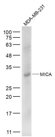
Sample:
MDA-MB-231(Human) Cell Lysate at 30 ug
Primary: Anti-MICA (SL0832R) at 1/500 dilution
Secondary: IRDye800CW Goat Anti-Rabbit IgG at 1/20000 dilution
Predicted band size: 43 kD
Observed band size: 33 kD
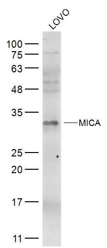
Sample:
LOVO(Human) Cell Lysate at 30 ug
Primary: Anti-MICA (SL0832R) at 1/500 dilution
Secondary: IRDye800CW Goat Anti-Rabbit IgG at 1/20000 dilution
Predicted band size: 43 kD
Observed band size: 33 kD
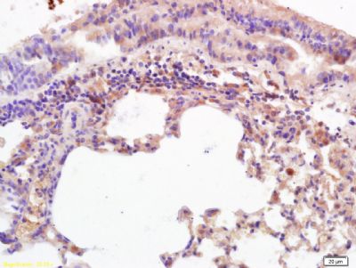
Tissue/cell: mouse lung tissue; 4% Paraformaldehyde-fixed and paraffin-embedded;
Antigen retrieval: citrate buffer ( 0.01M, pH 6.0 ), Boiling bathing for 15min; Block endogenous peroxidase by 3% Hydrogen peroxide for 30min; Blocking buffer (normal goat serum,SLC0005) at 37℃ for 20 min;
Incubation: Anti-MICA/MHC I a Polyclonal Antibody, Unconjugated(SL0832R) 1:200, overnight at 4°C, followed by conjugation to the secondary antibody(SP-0023) and DAB(SLC0010) staining
Tissue/cell: mouse intestine tissue; 4% Paraformaldehyde-fixed and paraffin-embedded;
Antigen retrieval: citrate buffer ( 0.01M, pH 6.0 ), Boiling bathing for 15min; Block endogenous peroxidase by 3% Hydrogen peroxide for 30min; Blocking buffer (normal goat serum,SLC0005) at 37℃ for 20 min;
Incubation: Anti-MICA/MHC I a Polyclonal Antibody, Unconjugated(SL0832R) 1:200, overnight at 4°C, followed by conjugation to the secondary antibody(SP-0023) and DAB(SLC0010) staining
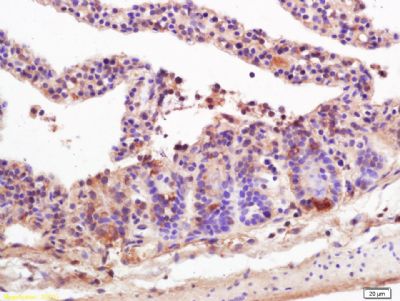
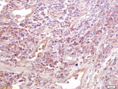
Tissue/cell: human lung carcinoma; 4% Paraformaldehyde-fixed and paraffin-embedded;
Antigen retrieval: citrate buffer ( 0.01M, pH 6.0 ), Boiling bathing for 15min; Block endogenous peroxidase by 3% Hydrogen peroxide for 30min; Blocking buffer (normal goat serum,SLC0005) at 37℃ for 20 min;
Incubation: Anti-MICA/MHC I a Polyclonal Antibody, Unconjugated(SL0832R) 1:200, overnight at 4°C, followed by conjugation to the secondary antibody(SP-0023) and DAB(SLC0010) staining
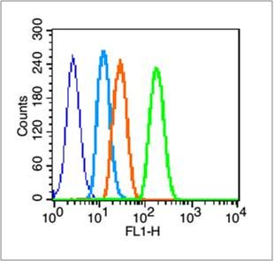
Blank control (blue line): A431 (blue).
Primary Antibody (green line): Rabbit Anti-MICA antibody (SL0832R)
Dilution: 1μg /10^6 cells;
Isotype Control Antibody (orange line): Rabbit IgG .
Secondary Antibody (white blue line): Goat anti-rabbit IgG-FITC
Dilution: 1μg /test.
Protocol
The cells were fixed with 2% paraformaldehyde (10 min) , then permeabilized with 90% ice-cold methanol for 30 min on ice. Cells stained with Primary Antibody for 30 min at room temperature. The cells were then incubated in 1 X PBS/2%BSA/10% goat serum to block non-specific protein-protein interactions followed by the antibody for 15 min at room temperature. The secondary antibody used for 40 min at room temperature. Acquisition of 20,000 events was performed.
|
|
|