[IF=6.038] Liao, Xiaodan. et al. Fullerene nanoparticles for the treatment of ulcerative colitis. 2021 Nov 02 WB ; Rat.
[IF=4.268] Jian-xia Wen. et al. Therapeutic Effects and Potential Mechanism of Dehydroevodiamine on N-Methyl-N′-Nitro-N-Nitrosoguanidine-Induced Chronic Atrophic Gastritis. Phytomedicine. 2021 Jun;:153619 WB ; Rat.
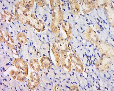
[IF=3.216] Yanhong Wang. et al. Intermedin Alleviates Renal Ischemia-Reperfusion Injury and Enhances Neovascularization in Wistar Rats. Drug Des Dev Ther. 2020; 14: 4825–4834 IHC ; Rat.
[IF=2.72] Jiebin Li. et al. Targeted Temperature Management Suppresses Hypoxia-Inducible Factor-1α and Vascular Endothelial Growth Factor Expression in a Pig Model of Cardiac Arrest. 2021 Jan 05 WB ; Pig.
[IF=3.923] Li H et al. siRNA‐mediated silencing of PAI‐1 gene acts as a promoter over the recanalization of endothelial progenitor cells in rats with venous thrombosis. J Cell Physiol. 2019 Apr 14. ICC ; Rat.
[IF=0] Wan et al. Endostatin, an angiogenesis inhibitor, ameliorates bleomycin-induced pulmonary fibrosis in rats. (2013) Respir.Re. 14:56 IHSLCP ; Rat.
[IF=2.02] Wang, Zheng, et al. "RhGH attenuates ischemia injury of intrahepatic bile ducts relating to liver transplantation." Journal of Surgical Research 171.1 (2011): 300-310. IHSLCP ; Rat.
[IF=2.84] Wei, Shuping, et al. "Targeted Contrast-Enhanced Ultrasound Imaging of Angiogenesis in an Orthotopic Mouse Tumor Model of Renal Carcinoma."Ultrasound in Medicine & Biology (2014) IHSLCP ; Mouse.
[IF=4.15] Wang, Nan, et al. "Vascular endothelial growth factor stimulates endothelial differentiation from mesenchymal stem cells via Rho/myocardin-related transcription factor-A signaling pathway." The International Journal of Biochemistry & Cell Biology (2013). WB ; Human, Rat.
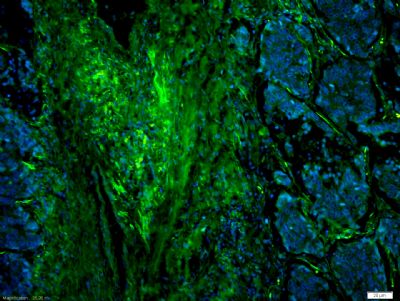
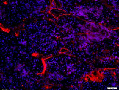
[IF=3.375] Zheng Chen. et al. Glycine attenuates cerebrovascular remodeling via glycine receptor alpha 2 and vascular endothelial growth factor receptor 2 after stroke. Am J Transl Res. 2020; 12(10): 6895–6907 IF,IHC ; Rat.
[IF=3.73] Gao, Jin-Hang, et al. "Celecoxib Ameliorates Portal Hypertension of the Cirrhotic Rats through the Dual Inhibitory Effects on the Intrahepatic Fibrosis and Angiogenesis." PLOS ONE 8.7 (2013): e69309. WB, IHSLCP ; Rat.
[IF=3.072] Xue Y et al. miR‐205‐5p inhibits psoriasis‐associated proliferation and angiogenesis: Wnt/β‐catenin and mitogen‐activated protein kinase signaling pathway are involved. J Dermatol
. 2020 Aug;47(8):882-892. WB ; Mouse&Human.
[IF=0.78] Yang K et al. Expression and distribution of HIF-1α, HIF-2α, VEGF, VEGFR-2 and HIMF in the kidneys of Tibetan sheep, Plain sheep and goat. Folia Morphol (Warsz). 2020 Feb 5. IHSLCP ; Sheep.
[IF=2.705] Liu Z et al. MRI analysis of hydrogel-loaded apatinib for local therapy of hepatocellular carcinoma model in nude mice. Biochem Biophys Res Commun. 2019 Feb 5;509(2):529-534. IHC ; Human.
[IF=1.77] Wang et al. Effects of minocycline on apoptosis and angiogenesis-related protein expression in a rat model of intracerebral hemorrhage. (2012) Neural.Regen.Res. 7:595-600 IHSLCF ; Rat.
[IF=1.069] Yang, Lei, et al. "Promotive effect of salvia extract on angiogenesis of myocardium in rats with myocardial infarction." Int J Clin Exp Med 10.6 (2017): 9245-9251. IHSLCF ; Rat.
[IF=3.17] Li, Junqin, et al. "Fatty Acid Synthase Mediates the Epithelial-Mesenchymal Transition of Breast Cancer Cells." International Journal of Biological Sciences 10.2 (2014): 171-36. WB ; Human.