[IF=9.776] Ruijing Zhao. et al. Inhalation of MSSLCEVs is a noninvasive strategy for ameliorating acute lung injury. J Control Release. 2022 May;345:214 FC ; Human.
[IF=2.984] Heli Zhang. et al. FN1 promotes chondrocyte differentiation and collagen production via TGF-β/PI3K/Akt pathway in mice with femoral fracture. Gene. 2021 Feb;769:145253 IHC ; Mouse.
[IF=4.432] Chunxia Chen. et al. Fracture repair by IOX2: Regulation of the hypoxia inducible factor-1α signaling pathway and BMSCs. Eur J Pharmacol. 2022 Apr;921:174864 IHC ; Rat.
[IF=5.923] Jiaqiang Deng. et al. Curcumin Alleviates the Senescence of Canine Bone Marrow Mesenchymal Stem Cells during In Vitro Expansion by Activating the Autophagy Pathway. Int J Mol Sci. 2021 Jan;22(21):11356 FC ; Dog.
[IF=3.647] Chunxia Chen. et al. HIF/Ca2+/NO/ROS is critical in roxadustat treating bone fracture by stimulating the proliferation and migration of BMSCs. Life Sci. 2021 Jan;264:118684 IHC ; Rat.
[IF=8.352] Chanjuan Dong. et al. Graphene-based conductive fibrous scaffold boosts sciatic nerve regeneration and functional recovery upon electrical stimulation. Appl Mater Today. 2020 Dec;21:100870 IHC ; Rat.
[IF=5.076] Feng T et al. Melatonin Protects Goat Spermatogonial Stem Cells against Oxidative Damage during Cryopreservation by Improving Antioxidant Capacity and Inhibiting Mitochondrial Apoptosis PathwayOxid Med Cell Longev.2020 Dec 31;2020:5954635. IF ; Goat.
[IF=8.579] Jipeng Jiang. et al. Implantation of regenerative complexes in traumatic brain injury canine models enhances the reconstruction of neural networks and motor function recovery. Theranostics. 2021; 11(2): 768–788 FC ; Human.
[IF=3.276] Wang Y et al. Establishment and Preclinical Therapy of Patient-derived Hepatocellular Carcinoma Xenograft Model.Immunol Lett. 2020 Jul;223:33-43. IHC ; human.
[IF=4.963] Jin J et al. Exosome secreted from adipose-derived stem cells attenuates diabetic nephropathy by promoting autophagy flux and inhibiting apoptosis in podocyte.Stem Cell Res Ther. 2019 Mar 15;10(1):95. ICF ; Human.
[IF=0.52] Utomo, Pamudji, et al. "Decreasing SDF1-CXCR4 Expression after Adipose-Derived Mesenchymal Stem Cells (ASCS) Treatment Combined with Freeze-Dried Amniotic Membrane Wrapping in Rat Sciatic Nerve Injury." International Journal of ChemTech Research. IF(ICC) ; Rat.
[IF=0] Gstraunthaler, Gerhard, et al. "Human platelet lysates successfully replace fetal bovine serum in adipose-derived adult stem cell culture." Journal of Advanced Biotechnology and Bioengineering 2.1 (2014): 1-11. IF(ICC) ; Human.
[IF=1.43] Zeng, Biao, et al. "Increased expression of importin13 in endometriosis and endometrial carcinoma." Medical Science Monitor 18.6 (2012): CR361-CR367. IHSLCP ; Human.
[IF=2.26] Yu, Xian-huan, et al. "Clinicopathological characteristics of 20 cases of hepatocellular carcinoma with bile duct tumor thrombi." Digestive diseases and sciences 56.1 (2011): 252-259. IHSLCP ; Human.
[IF=4.648] Long et al. Mash1-dependent Notch Signaling Pathway Regulates GABAergic Neuron-Like Differentiation from Bone Marrow-Derived Mesenchymal Stem Cells. (2017) Aging.Di. 8:301-313 FCM ; Rat.
[IF=3.427] Gao et al. Common expression of stemness molecular markers and early cardiac transcription factors in human Wharton's jelly-derived mesenchymal stem cells and embryonic stem cells. (2013) Cell.Transplan. 22:1883-900 FCM ; Human.
[IF=1.977] Jianqiang Zhao. et al. In vitro facilitating role of polygonatum sibiricum polysaccharide in osteogenic differentiation of bone marrow mesenchymal stem cells from patients with multiple myeloma. 2021 Apr 23 FC ; Human.
[IF=2.94] Wang, Kai, et al. "Over-expression of Mash1 improves the GABAergic differentiation of bone marrow mesenchymal stem cells< i> in vitro." Brain Research Bulletin (2013). FCM ; Rat.
[IF=0.918] Chen F et al.
The biological characteristics of sheep umbilical cord mesenchymal stem cells.Can J Vet Res. 2018 Jul;82(3):216-224. ICF ; lamb.
[IF=2.634] Ma C et al. Identification and Multilineage Potential Research of a Novel Type of Adipose-Derived Mesenchymal Stem Cells from Goose Inguinal Groove. DNA Cell Biol. 2018 Sep;37(9):731-741. ICF ; Goose.
[IF=4.034] Wang YL et al. Preinduction with bone morphogenetic protein-2 enhances cardiomyogenic differentiation of c-kit+ mesenchymal stem cells and repair of infarcted myocardium.Int J Cardiol. 2018 Aug 15;265:173-36. FCM ; Rat.
[IF=1.461] Pei W et al. Biological characterization and pluripotent identification of ovine amniotic fluid stem cells. Cytotechnology. 2018 Jun;70(3):1009-1021. IF&FCM ; ovine embryo.
[IF=2.66] Li et al. Perichondrium mesenchymal stem cells inhibit the growth of breast cancer cells via the DKK-1/Wnt/β-catenin signaling pathway. (2016) Oncol.Rep. 36:936-44 IF(ICC) ; Rat.
[IF=1.92] Ma, Caiyun, et al. "Cryopreservation and multipotential characteristics evaluation of a novel type of mesenchymal stem cells derived from Small Tailed Han Sheep fetal lung tissue." Cryobiology (2017). FCM ; Sheep.
[IF=1.06] Scott, Erin M., et al. "Early histopathologic changes in the retina and optic nerve in canine primary angle‐closure glaucoma." Veterinary ophthalmology16.s1 (2013): 79-86. IHSLCP ; Dog.
[IF=1.8] Vansandt, Lindsey M., et al. "Conservation of spermatogonial stem cell marker expression in undifferentiated felid spermatogonia." Theriogenology(2016). IHSLCP ; Other Species.
[IF=3.36] Long, Q., et al. "Bone marrow mesenchymal stem cell transplantation improves cognitive impairment via up-regulation of hippocampal GABAergic system in a rat model of chronic cerebral hypoperfusion." Neuroscience (2015). Rat.
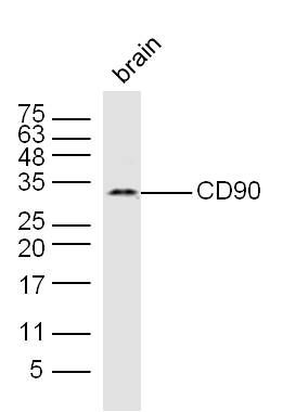
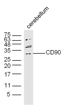
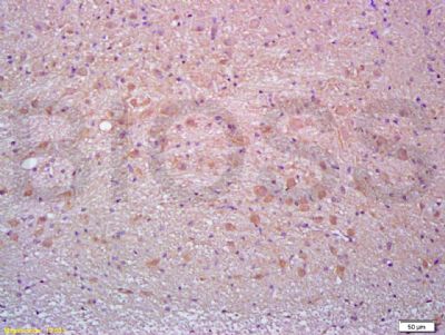
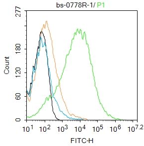
[IF=1.52] Tepekoy, Filiz, et al. "CD90 and CD105 expression in the mouse ovary and testis at different stages of postnatal development." Reproductive Biology(2015). IHSLCP ; Mouse.
[IF=5.9] Chen, Guobao, et al. "3D Scaffolds with Different Stiffness but Same Microstructure for Bone Tissue Engineering." ACS Applied Materials & Interfaces (2015). IHSLCF ; Rat.
[IF=2.88] Long, Qianfa, et al. "Genetically engineered bone marrow mesenchymal stem cells improve functional outcome in a rat model of epilepsy." Brain Research (2013). Rat.