[IF=1.88] Shihe Zhang. et al. Astragalus polysaccharide regulates brown adipocytes differentiation by miR-6911 targeting Prdm16. 2021 Nov 05 WB ; Mouse.
[IF=3.105] Huan-Tong Wu. et al. Edaravone attenuates H2O2 or glutamate-induced toxicity in hippocampal neurons and improves AlCl3/D-galactose induced cognitive impairment in mice. Neurotoxicology. 2021 Jul;85:68 WB ; Rat.
[IF=4.357] Li et al. Morusin suppresses breast cancer cell growth in vitro and in vivo through C/EBPβ and PPARγ mediated lipoapoptosis. (2015) J.Exp.Clin.Cancer.Res. 34:137 WB ; Human.
[IF=5.62] He, Ting, et al. "Tumor cell-secreted angiogenin induces angiogenic activity of endothelial cells by suppressing miR-542-3p." Cancer Letters (2015). WB ; Human.
[IF=1.922] Wang F et al. Insulin‑like growth factor I promotes adipogenesis in hemangioma stem cells from infantile hemangiomas. Mol Med Rep. 2019 Jan 24. WB ; Human.
[IF=4.575] Ren, Teng-Teng. et al. Gisenoside Rg1 attenuates cadmium-induced neurotoxicity in vitro and in vivo by attenuating oxidative stress and inflammation. Inflamm Res. 2021 Dec;70(10):1151-1164 WB ; Mouse.
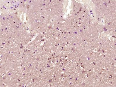
[IF=2.02] Jiejuan Lai. et al. Comparison of the biological and functional characteristics of mesenchymal stem cells from intrahepatic and identical bone marrow. Stem Cell Res. 2021 Aug;55:102477 IHC ; Mouse.
[IF=4.259] Wu Z et al. Co-infection of Mycoplasma gallisepticum and Escherichia coli Triggers Inflammatory Injury Involving the IL-17 Signaling Pathway. Front Microbiol. 2019 Nov 15;10:2615. WB ; Chicken.
[IF=1.832] Zhang T et al. Dietary Sea Buckthorn Pomace Induces Beige Adipocyte Formation in Inguinal White Adipose Tissue in Lambs. Animals (Basel). 2019 Apr 24;9(4). pii: E193. WB ; ram lambs.
[IF=1.448] Zhang K et al. Insulin‑like growth factor 2 promotes the adipogenesis of hemangioma‑derived stem cells. Exp Ther Med. 2019 Mar;17(3):1663-1669. WB ; Human.