[IF=5.753] Chaohua Cong. et al. PAK4 suppresses motor neuron degeneration in hSOD1G93A‐linked amyotrophic lateral sclerosis cell and rat models. 2021 Feb 21 IF,IHC ; Mouse.
[IF=5.878] Lin Zhu. et al. Neuroprotective effects of salidroside on ageing hippocampal neurons and naturally ageing mice via the PI3K/Akt/TERT pathway. 2021 Aug 09 WB ; Rat.
[IF=13.281] Yu Hei. et al. Multifunctional Immunoliposomes Enhance the Immunotherapeutic Effects of PD-L1 Antibodies against Melanoma by Reprogramming Immunosuppressive Tumor Microenvironment. 2021 Dec 16 IF,IHC ; Mouse.
[IF=6.291] Hongmei Zhou. et al. Downregulation of beclin 1 restores arsenite-induced impaired autophagic flux by improving the lysosomal function in the brain. Ecotox Environ Safe. 2022 Jan;229:113066 IF ; Mouse.
[IF=5.75] Zhifei Wang. et al. Human Cytomegalovirus Immediate Early Protein 2 Protein Causes Cognitive Disorder by Damaging Synaptic Plasticity in Human Cytomegalovirus-UL122-Tg Mice. Front Aging Neurosci. 2021; 13: 720582 IF,IHC ; Mouse.
[IF=2.276] Zhang Heng. et al. Bone Morphogenetic Protein-7 (BMP-7) Promotes Neuronal Differentiation of Bone Marrow Mesenchymal Stem Cells (BMSCs) In Vitro. Biomed Res Int. 2021;2021:7239783 WB,IF,ICC ; Rat.
[IF=3.414] Wang K et al. Harpagide from Scrophularia protects rat cortical neurons from oxygen-glucose deprivation and reoxygenation-induced injury by decreasing endoplasmic reticulum stress. J Ethnopharmacol. 2020 Jan 31;253:112614. WB ; Rat.
[IF=3.241] Che H et al. EPA-Enriched Ethanolamine Plasmalogen and EPA-Enriched Phosphatidylethanolamine Enhance BDNF/TrkB/CREB Signaling and inhibit Neuronal Apoptosis in vitro and in vivo. Food Funct. 2020 Feb 11. ICF ; Rat.
[IF=2.772] Zhou J et al. Imbalance of Microglial TLR4/TREM2 in LPS-Treated APP/PS1 Transgenic Mice: A Potential Link Between Alzheimer's Disease and Systemic Inflammation Neurochemical Research.2019. TUNEL ; Mouse.
[IF=3.97] Lu, Lihua, et al. "AMP-activated protein kinase activation in mediating phenylalanine-induced neurotoxicity in experimental models of phenylketonuria." Journal of inherited metabolic disease (2017): 1-9. WB ; Mouse.
[IF=1.61] Usui, Tatsuya, et al. "Establishment of a novel three‐dimensional primary culture model for hippocampal neurogenesis." Physiological Reports 5.12 (2017): e13318. IF(IHSLCP) ; Mouse.
[IF=3.105] Shanshan Zhang. et al. Pregnancy exposure to carbon black nanoparticles induced neurobehavioral deficits that are associated with altered m6A modification in offspring. Neurotoxicology. 2020 Dec;81:40 WB ; Mouse.
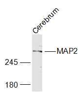
[IF=5.391] Jiang Y et al. A mutation in MAP2 is associated with prenatal hair follicle density. ASEB J. 2019 Dec;33(12):14479-14490. IHSLCP ; Pig.
[IF=5.16] Yang YQ et al. Wild-type p53-induced phosphatase 1 down-regulation promotes apoptosis by activating the DNA damage-response pathway in amyotrophic lateral sclerosis. Neurobiol Dis. 2019 Oct 30;134:104648. IHF ; Mouse.
[IF=3.265] Zhang Y et al. Exposure to carbon black nanoparticles during pregnancy persistently damages the cerebrovascular function in female mice. Toxicology. 2019 Apr 22;422:44-52. IHF&WB ; Mouse.
[IF=3.076] Tang Q et al. Ferroptosis is newly characterized form of neuronal cell death in response to arsenite exposure.Neurotoxicology. 2018 Jul;67:27-36. IF ; Mouse.
[IF=2.3] Wang, Jin, et al. "Neuroprotective Effects of Wnt/ß-catenin signaling pathway against Aβ-induced Tau protein over-phosphorylation in PC12 cells."Biochemical and Biophysical Research Communications (2016). Rat.
[IF=2.65] Zuo, Daiying, et al. "Existence of glia mitigated ketamine-induced neurotoxicity in neuron-glia mixed cultures of neonatal rat cortex and the glia-mediated protective effect of 2-PMPA." Neurotoxicology (2014). Rat.
[IF=4.35] Gu, LiZe, et al. "Early activation of nSMase2/ceramide pathway in astrocytes is involved in ischemia-associated neuronal damage via inflammation in rat hippocampi." Journal of Neuroinflammation 10.1 (2013): 109. Rat.