[IF=2.611] Cui, Yan. et al. Effects of PHD and HSP90 on erythropoietin production in yak (Bos grunniens) renal interstitial fibroblast-like cells under hypoxia. J Mol Histol. 2022 Jan;:1-17 IHC ; Yak.
[IF=2.752] Yanyu He. et al. Altered Hypoxia-Induced and Heat Shock Protein Immunostaining in Secondary Hair Follicles Associated with Changes in Altitude and Temperature in Tibetan Cashmere Goats. Animals-Basel. 2021 Oct;11(10):2798 IF,IHC ; goat.
[IF=1.832] He Y et al. Association of Age with the Expression of Hypoxia-Inducible Factors HIF-1α, HIF-2α, HIF-3α and VEGF in Lung and Heart of Tibetan Sheep. Animals (Basel). 2019 Sep 11;9(9). pii: E673. IHSLCP ; Sheep.
[IF=1.58] Li, Hao, et al. "Prognostic value of CD147 and HIF-2α expression in localized clear cell renal cell carcinoma." Int J Clin Exp Pathol 9.9 (2016): 9394-9400. IHSLCP ; Human.
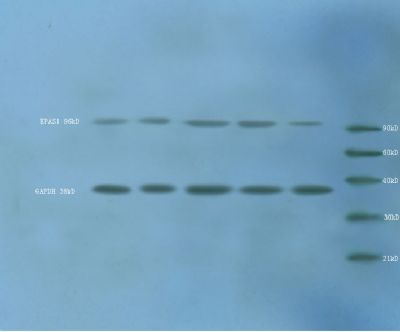
[IF=3.35] Li, Xu, et al. "Oxidative stress induces the decline of brain EPO expression in aging rats." Experimental Gerontology (2016). WB ; Rat.
[IF=0.78] Yang K et al. Expression and distribution of HIF-1α, HIF-2α, VEGF, VEGFR-2 and HIMF in the kidneys of Tibetan sheep, Plain sheep and goat. Folia Morphol (Warsz). 2020 Feb 5. IHSLCP ; Sheep.
[IF=2.466] Jinjiang Yang. et al. Platelet-rich plasma attenuates interleukin-1β-induced apoptosis and inflammation in chondrocytes through targeting hypoxia-inducible factor-2α. Tissue Cell. 2021 Sep;:101646 WB ; rat.
[IF=4.451] Shiying Huang. et al. Jujube polysaccharides mitigated anemia in rats with chronic kidney disease: Regulation of short chain fatty acids release and erythropoietin production. J Funct Foods. 2021 Nov;86:104673 IHC ; Rat.
[IF=5.174] Xiong Y et al. Physiological and genetic convergence supports hypoxia resistance in high-altitude songbirdsPLoS Genet.2020 Dec 28;16(12):e1009270. WB ; Bird.
[IF=3.414] Tang Q et al. Aqueous extract from You-Gui-Yin ameliorates cognitive impairment of chronic renal failure micethrough targeting hippocampal CaMKIIα/CREB/BDNF and EPO/EPOR pathways. J Ethnopharmacol. 2019 Jul 15;239:111925. WB ; Mouse.
[IF=1.719] Lixin L et al. Effect of Hypoxia on the Muscle Fiber Switching Signal Pathways CnA/NFATc1 and Myostatin in Mouse Myocytes. Acta Histochem. 2019 Jul;121(5):539-545. WB ; Mouse.