[IF=3.159] Wan Boyang. et al. Zearalenone promotes follicle development through activating SIRT1/PGSLC1α signaling pathway in the ovaries of weaned gilts. J Anim Sci. 2022 Feb;: WB ; Pig(Gilt).
[IF=2.303] Suihui Li. et al. Hsa_circ_0048674 facilitates hepatocellular carcinoma progression and natural killer cell exhaustion depending on the regulation of miR-223-3p/PDL1. Histol Histopathol. 2022 Feb 21;1888 WB ; Human.
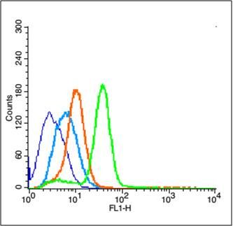
[IF=5.81] Yu TT. et al. Chlorin e6-Induced Photodynamic Effect Polarizes the Macrophage Into an M1 Phenotype Through Oxidative DNA Damage and Activation of STING.. Front Pharmacol. 2022 Mar;13:837784-837784 WB ; Mouse.
[IF=4.432] Ning Han. et al. Ferroptosis triggered by dihydroartemisinin facilitates chlorin e6 induced photodynamic therapy by inhibiting GPX4 and enhancing ROS. Eur J Pharmacol. 2022 Feb;:174797 WB ; Mouse.
[IF=4.553] Yu, Ting-Ting. et al. Harnessing chlorin e6 loaded by functionalized iron oxide nanoparticles linked with glucose for target photodynamic therapy and improving of the immunogenicity of lung cancer. J Cancer Res Clin. 2022 Jan;:1-13 WB,IF,IHC ; Mouse.
[IF=4.546] Qing Li. et al. Fumonisin B1 Inhibits Cell Proliferation and Decreases Barrier Function of Swine Umbilical Vein Endothelial Cells. Toxins. 2021 Dec;13(12):863 WB ; Pig.
[IF=3.743] Wang C et al. 4-Amino-2-trifluoromethyl-phenyl retinate induced differentiation of human myelodysplastic syndromes SKM-1 cell lines by up-regulating DDX23. Biomed Pharmacother. 2019 Dec 16;123:109736. WB ; Human.
[IF=8.402] Zhang,et al.Lysosomal deposition of copper oxide nanoparticles triggers HUVEC cells death.(2018) Biomaterials. 161:228-239. WB ; Human.
[IF=5.878] Jolly,et al.Targeted endothelial gene deletion of Triggering Receptor Expressed on Myeloid cells-1 protects mice during septic shock.(2018) Cardiovascular Research. 114:907-918. IF(IHSLCF) + IF(ICC) ; Mouse + Human.
[IF=2.34] Zhou et al. Induced pluripotent stem cell-conditioned medium suppresses pulmonary fibroblast-to-myofibroblast differentiation via the inhibition of TGF-β1/Smad pathway. (2018) Int.J.Mol.Med. 41:473-484 WB ; Human.
[IF=1.84] Zhao, Yong, et al. "Inhibition of peripubertal sheep mammary gland development by cysteamine through reducing progesterone and growth factor production." Theriogenology (2016). WB ; Sheep.
[IF=1.837] Yang Zhang. et al. Plumbagin Inhibits Proliferation, Migration, and Invasion of Retinal Pigment Epithelial Cells Induced by FGF-2. Tissue Cell. 2021 Oct;72:101547 WB ; Human.
[IF=0.439] Wu Z et al. Compound xiebai capsule alleviates pulmonary vascular remodeling in monocrotaline-induced pulmonary arterial hypertension in rats. Tropical Journal of Pharmaceutical Research January 2020; 19 (1): 107-114. WB ; Rat.
[IF=5.808] Jin J et al. A novel S1P1 modulator IMMH002 ameliorates psoriasis in multiple animal models. Acta Pharmaceutica Sinica B. 2019. IHSLCP ; Mouse.
[IF=3.457] Gao Y et al. Ginsenoside Re inhibits vascular neointimal hyperplasia in balloon-injured carotid arteries through activating the eNOS/NO/cGMP pathway in rats
Y Gao, CY Gao, P Zhu, SF Xu, YM Luo, J Deng… - Biomedicine & …, 2018Biomed Pharmacother. 2018 Oct;106:1091-1097. IHSLCP ; Rat.
[IF=6.375] Zhou,et al.CXCR4 antagonist AMD620 enhances the response of MDA-MB-231 triple-negative breast cancer cells to ionizing radiation.(2018) Cancer Letters. 418:196-203. IHSLCP + WB ; Mouse.
[IF=2.78] Chai et al. Hypoxia induces pulmonary arterial fibroblast proliferation, migration, differentiation and vascular remodeling via the PI3K/Akt/p70S6K signaling pathway. (2018) Int.J.Mol.Med. 41:2461-2472 WB ; Rat.
[IF=1.69] Ji, Wei, et al. "Triptolide inhibits proliferation, differentiation and induces apoptosis of osteoblastic MC3T3‑E1 cells." Molecular Medicine Reports. WB ; Mouse.
[IF=5.01] Liang, Sixian, et al. "Silencing of CXCR4 sensitizes triple-negative breast cancer cells to cisplatin." Oncotarget 6.2 (2015): 1020-1030. IHSLCP ; Mouse.