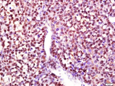
[IF=1.066] Zong-Ding Wang. et al. A Novel Hepatectomy Model in Mice Using a Gutta Cutter Tool: A Feasibility Study and Preliminary Results. Transpl P. 2022 Mar;: IHC ; Mouse.
[IF=5.279] Yan Zhang. et al. Ganoderic Acid A To Alleviate Neuroinflammation of Alzheimer’s Disease in Mice by Regulating the Imbalance of the Th17/Tregs Axis. J Agr Food Chem. 2021;69(47):14204–14214 WB ; Mouse.
[IF=5.037] Hongwu Meng. et al. Interleukin-9 attenuates inflammatory response and hepatocyte apoptosis in alcoholic liver injury. Life Sci. 2022 Jan;288:12036 WB ; Mouse.
[IF=4.105] Ai Jin. et al. TSLP-induced collagen type-I synthesis through STAT3 and PRMT1 is sensitive to calcitriol in human lung fibroblasts. Bba-Mol Cell Res. 2021 Jun;:119083 WB ; Human.
[IF=2.886] Yusi Liu. et al. Synergistic Effects of Resveratrol and Temozolomide Against Glioblastoma Cells: Underlying Mechanism and Therapeutic Implications. Cancer Manag Res. 2020; 12: 8341–8354 ICC ; Rat, Human.
[IF=2.05] Zhang M et al. Hepatoprotective effect and possible mechanism of phytoestrogen calycosin on carbon tetrachloride–induced liver fibrosis in mice. Naunyn Schmiedebergs Arch Pharmacol
. 2020 May 30. WB ; Mouse.
[IF=2.736] Hao Y et al. IL-6/STAT3 mediates the HPV18 E6/E7 stimulated upregulation of MALAT1 gene in cervical cancer HeLa cells. Virus Res. 2020 May;281:197907. WB ; human.
[IF=4.868] Wang G et al. Protective Effect of Methane-Rich Saline on Acetic Acid-Induced Ulcerative Colitis via Blockingthe TLR4/NF-κB/MAPK Pathway and Promoting IL-10/JAK1/STAT3-Mediated Anti-inflammatory Response. Oxid Med Cell Longev. 2019 Apr 28;2019:7850324. WB ; Mouse.
[IF=3.414] Zhou H et al. Paris saponin VII extracted from trillium tschonoskii suppresses proliferation and induces apoptosis of human colorectal cancer cells. J Ethnopharmacol. 2019 Jul 15;239:111903. WB ; Human.
[IF=2.81] Badin et al. Muscle Arnt/Hif1β Is Dispensable in Myofiber Type Determination, Vascularization and Insulin Sensitivity. (2016) PLoS.One. 11:e0168457 WB ; Mouse.
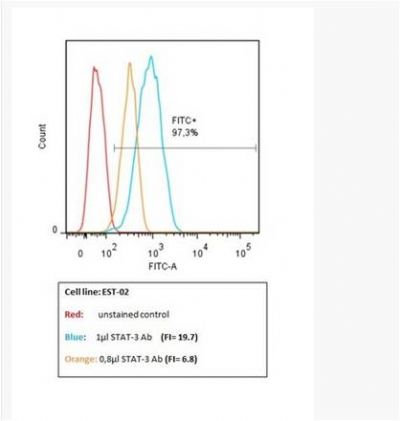
[IF=3.36] Iriyama et al. Direct effect of dasatinib on signal transduction pathways associated with a rapid mobilization of cytotoxic lymphocytes. (2016) Cancer.Med. 5:3223-3234 FC/FACS ; Human.
[IF=2.82] Gao, Ling, et al. "Morusin shows potent antitumor activity for human hepatocellular carcinoma in vitro and in vivo through apoptosis induction and angiogenesis inhibition." Drug Design, Development and Therapy 11 (2017): 1789. WB ; Human.
[IF=2.991] Yang L et al. 15-Lipoxygenase-2/15(S)-hydroxyeicosatetraenoic acid regulates cell proliferation and metastasis via the STAT3 pathway in lung adenocarcinoma. Prostaglandins Other Lipid Mediat. 2018 Sep;138:31-40. WB&IHSLCP ; Human.
[IF=2.705] Li B et al. Increased hepcidin in hemorrhagic plaques correlates with iron-stimulated IL-6/STAT3 pathway activation in macrophages. Biochem Biophys Res Commun. 2019 Jul 23;515(2):394-400. IHC,WB ; Rabbit,Human&Mouse.
[IF=3.269] Leiming Sun. et al. Isoliquiritigenin attenuates acute renal injury through suppressing oxidative stress, fibrosis and JAK2/STAT3 pathway in streptozotocin-induced diabetic rats. Bioengineered. 2021;12(2):11188-1240 WB ; Rat.
[IF=3.347] Yuying Li. et al. Anthraquinone derivative C10 inhibits proliferation and cell cycle progression in colon cancer cells via the Jak2/Stat3 signaling pathway. Toxicol Appl Pharm. 2021 Mar;:115481 IF ; Human.
[IF=4.187] Jiajia Wang. et al. TAK-242 ameliorates DSS-induced colitis by regulating the gut microbiota and the JAK2/STAT3 signaling pathway. Microb Cell Fact. 2020 Dec;19(1):1-17 WB ; Mouse.
[IF=2.05] Mengmeng Zhang. et al. Hepatoprotective effect and possible mechanism of phytoestrogen calycosin on carbon tetrachloride–induced liver fibrosis in mice. N-S Arch Pharmacol. 2021 Jan;394(1):189-204 WB ; Mouse.
[IF=3.606] Xuejiao Zhou. et al. The novel ALK inhibitor ZX‐29 induces apoptosis through inhibiting ALK and inducing ROS‐mediated endoplasmic reticulum stress in Karpas299 cells. 2020 Nov 02 WB ; Human.
[IF=2.51] Yang, Hai Li, et al. "Effect of suppressor of cytokine signaling 2 (SOCS2) on fat metabolism induced by growth hormone (GH) in porcine primary adipocyte." Molecular biology reports 39.9 (2012): 9113-9122. WB ; Pig.
[IF=8.724] Yong Tang. et al. Phosphorylation inhibition of protein-tyrosine phosphatase 1B tyrosine-152 induces bone regeneration coupled with angiogenesis for bone tissue engineering. Bioact Mater. 2021 Jul;6:2039 WB ; Mouse.
[IF=1.947] Gao M et al. Activating the interleukin-6-Gp130-STAT3 pathway ameliorates ventricular electrical stability in myocardial infarction rats by modulating neurotransmitters in the paraventricular nucleus. BMC Cardiovasc Disord. 2020 Feb 5;20(1):60. IHSLCP&WB ; Rat.
[IF=2.25] Li H et al. MicroRNA-204-5p suppresses IL6-mediated inflammatory response and chemokine generation in HK-2 renal tubular epithelial cells by targeting IL6R. Biochem Cell Biol. 2019 Apr;97(2):109-117. WB ; Human.
[IF=3.457] Ling L et al. MicroRNA-30e promotes hepatocyte proliferation and inhibits apoptosis in cecal ligation and puncture-induced sepsis through the JAK/STAT signaling pathway by binding to FOSL2.Biomed Pharmacother. 2018 Aug;104:411-419. WB ; Rat.
[IF=2.559] Bi J et al. PPARγ alleviated hepatocyte steatosis through reducing SOCS3 by inhibiting JAK2/STAT3 pathway. Biochem Biophys Res Commun. 2018 Apr 15;498(4):1037-1044. ICC&WB ; Rat.
[IF=0.9] Zhang, Yu, et al. "JAK-STAT signaling regulation of chicken embryonic stem cell differentiation into male germ cells." In Vitro Cellular & Developmental Biology-Animal (2017): 1-16. WB ; Chicken.
[IF=3.23] Sur, Swastika, et al. "Increased Expression of Phosphorylated Polo-Like Kinase 1 and Histone in Bypass Vein Graft and Coronary Arteries following Angioplasty." PloS one 11.1 (2016): e0147937. IF(IHSLCP) ; Pig.
[IF=3.23] Lee E-J, Lee SJ, Kim J-H, Kim K-J, Yang S-H, Jeong K-Y, et al. (2016) Radiation Inhibits Interleukin-12 Production via Inhibition of SLCRel through the Interleukin-6/ Signal Transducer and Activator of Transcription 3 Signaling Pathway in Dendritic Cells. PLoS ONE 11(1): e0146463. WB ; Mouse.