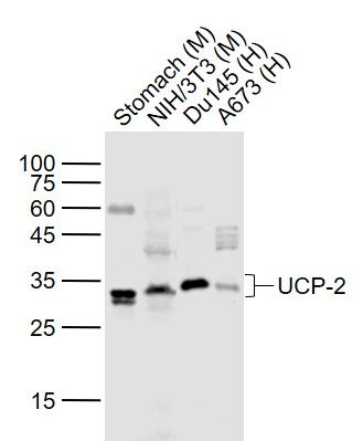
Sample:
Lane 1: Stomach (Mouse) Lysate at 40 ug
Lane 2: NIH/3T3 (Mouse) Cell Lysate at 30 ug
Lane 3: Du145 (Human) Cell Lysate at 30 ug
Lane 4: A673 (Human) Cell Lysate at 30 ug
Primary:
Anti-UCP-2 (SL1926R) at 1/1000 dilution
Secondary: IRDye800CW Goat Anti-Rabbit IgG at 1/20000 dilution
Predicted band size: 33 kD
Observed band size: 33 kD
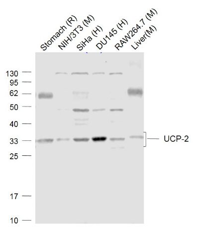
Sample:
Lane 1: Stomach (Rat) Lysate at 40 ug
Lane 2: NIH/3T3 (Mouse) Cell Lysate at 30 ug
Lane 3: SiHa (Human) Cell Lysate at 30 ug
Lane 4: DU145 (Human) Cell Lysate at 30 ug
Lane 5: RAW264.7 (Mouse) Cell Lysate at 30 ug
Lane 6: Liver (Mouse) Lysate at 40 ug
Primary: Anti-UCP-2 (SL1926R) at 1/1000 dilution
Secondary: IRDye800CW Goat Anti-Rabbit IgG at 1/20000 dilution
Predicted band size: 33 kD
Observed band size: 33 kD
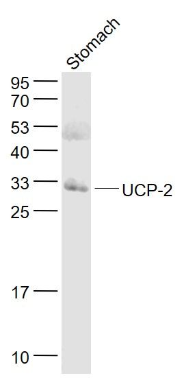
Sample:
Stomach (Mouse) Lysate at 40 ug
Primary: Anti- UCP-2 (SL1926R) at 1/1000 dilution
Secondary: IRDye800CW Goat Anti-Rabbit IgG at 1/20000 dilution
Predicted band size: 34 kD
Observed band size: 32 kD
Sample:
BSLV2(Mouse) Cell Lysate at 30 ug
Primary: Anti- UCP-2 (SL1926R) at 1/1000 dilution
Secondary: IRDye800CW Goat Anti-Rabbit IgG at 1/20000 dilution
Predicted band size: 34 kD
Observed band size: 32 kD
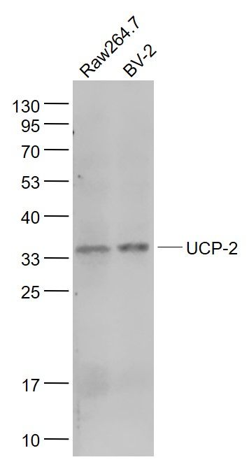
Sample:
Raw264.7(Mouse) Cell Lysate at 30 ug
BSLV2(Mouse) Cell Lysate at 30 ug
Primary: Anti- UCP-2 (SL1926R) at 1/500 dilution
Secondary: IRDye800CW Goat Anti-Rabbit IgG at 1/20000 dilution
Predicted band size: 34 kD
Observed band size: 34 kD
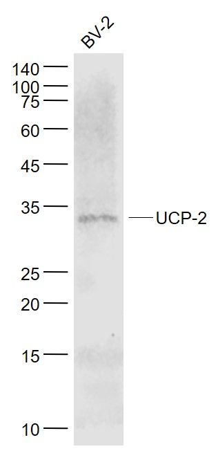
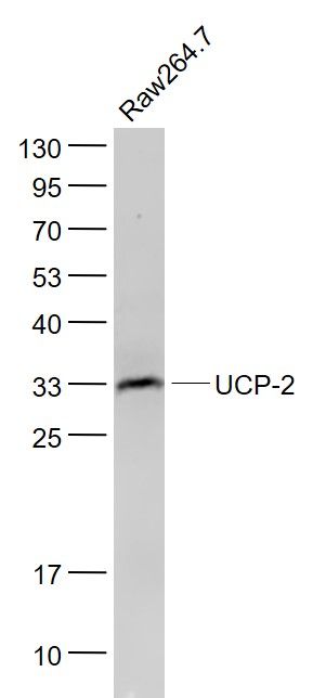
Sample:
Raw264.7(Mouse) Cell Lysate at 30 ug
Primary: Anti- UCP-2 (SL1926R) at 1/500 dilution
Secondary: IRDye800CW Goat Anti-Rabbit IgG at 1/20000 dilution
Predicted band size: 34 kD
Observed band size: 33 kD
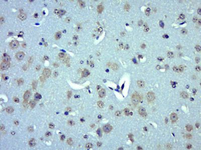
Paraformaldehyde-fixed, paraffin embedded (Mouse brain); Antigen retrieval by boiling in sodium citrate buffer (pH6.0) for 15min; Block endogenous peroxidase by 3% hydrogen peroxide for 20 minutes; Blocking buffer (normal goat serum) at 37°C for 30min; Antibody incubation with (UCP-2) Polyclonal Antibody, Unconjugated (SL1926R) at 1:400 overnight at 4°C, followed by operating according to SP Kit(Rabbit) (sp-0023) instructionsand DAB staining.
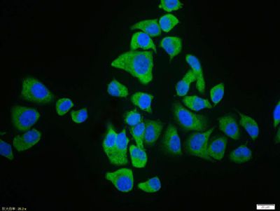
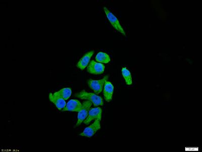
Hela cell; 4% Paraformaldehyde-fixed; Triton X-100 at room temperature for 20 min; Blocking buffer (normal goat serum, SLC0005) at 37°C for 20 min; Antibody incubation with (UCP-2) polyclonal Antibody, Unconjugated (SL1926R) 1:100, 90 minutes at 37°C; followed by a conjugated Goat Anti-Rabbit IgG antibody at 37°C for 90 minutes, DAPI (blue, C02-04002) was used to stain the cell nuclei.
Hela cell; 4% Paraformaldehyde-fixed; Triton X-100 at room temperature for 20 min; Blocking buffer (normal goat serum, SLC0005) at 37°C for 20 min; Antibody incubation with (UCP-2) polyclonal Antibody, Unconjugated (SL1926R) 1:100, 90 minutes at 37°C; followed by a conjugated Goat Anti-Rabbit IgG antibody at 37°C for 90 minutes, DAPI (blue, C02-04002) was used to stain the cell nuclei.
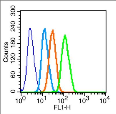
Blank control (blue line): Hela (blue).
Primary Antibody (green line): Rabbit Anti-UCP-2 antibody (SL1926R)
Dilution: 1μg /10^6 cells;
Isotype Control Antibody (orange line): Rabbit IgG .
Secondary Antibody (white blue line): F(ab’)2 fragment goat anti-rabbit IgG-FITC
Dilution: 1μg /test.
Protocol
The cells were fixed with 2% paraformaldehyde (10 min) , then permeabilized with 90% ice-cold methanol for 30 min on ice.Cells stained with Primary Antibody for 30 min at room temperature. The cells were then incubated in 1 X PBS/2%BSA/10% goat serum to block non-specific protein-protein interactions followed by the antibody for 15 min at room temperature. The secondary antibody used for 40 min at room temperature. Acquisition of 20,000 events was performed.
|