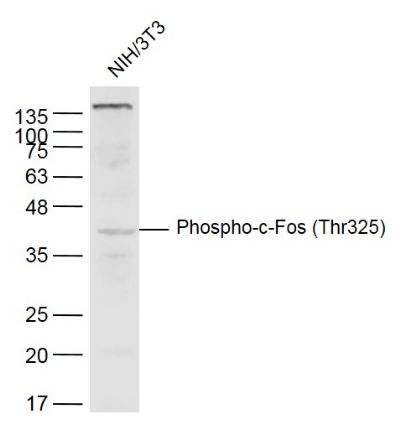
Sample:
NIH/3T3(Mouse) Cell Lysate at 40 ug
Primary: Anti-Phospho-c-Fos (Thr325) (SL3154R) at 1/300 dilution
Secondary: IRDye800CW Goat Anti-Rabbit IgG at 1/20000 dilution
Predicted band size: 41 kD
Observed band size: 41 kD
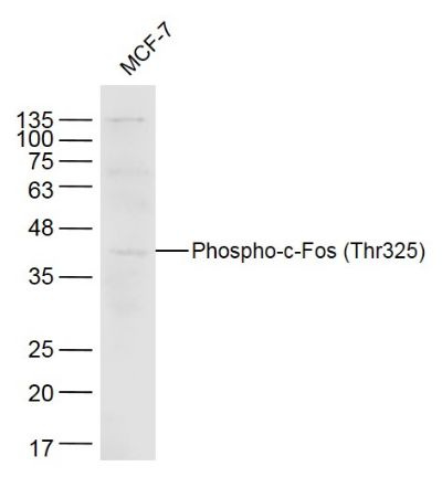
Sample:
MCF-7(Human)Cell Lysate at 40 ug
Primary: Anti-Phospho-c-Fos (Thr325) (SL3154R) at 1/300 dilution
Secondary: IRDye800CW Goat Anti-Rabbit IgG at 1/20000 dilution
Predicted band size: 41 kD
Observed band size: 41 kD
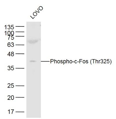
Sample:
LOVO(Human) Cell Lysate at 40 ug
Primary: Anti-Phospho-c-Fos (Thr325) (SL3154R) at 1/300 dilution
Secondary: IRDye800CW Goat Anti-Rabbit IgG at 1/20000 dilution
Predicted band size: 41 kD
Observed band size: 41 kD
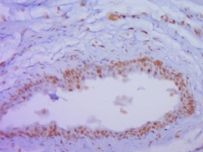
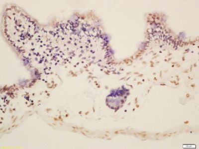
Paraformaldehyde-fixed, paraffin embedded (Rat urinary bladder); Antigen retrieval by boiling in sodium citrate buffer (pH6.0) for 15min; Block endogenous peroxidase by 3% hydrogen peroxide for 20 minutes; Blocking buffer (normal goat serum) at 37°C for 30min; Antibody incubation with (P-c-Fos (Thr325)) Polyclonal Antibody, Unconjugated (SL3154R) at 1:400 overnight at 4°C, followed by operating according to SP Kit(Rabbit) (sp-0023) instructionsand DAB staining.
Tissue/cell: mouse intestine tissue; 4% Paraformaldehyde-fixed and paraffin-embedded;
Antigen retrieval: citrate buffer ( 0.01M, pH 6.0 ), Boiling bathing for 15min; Block endogenous peroxidase by 3% Hydrogen peroxide for 30min; Blocking buffer (normal goat serum,SLC0005) at 37℃ for 20 min;
Incubation: Anti-Phospho-c-Fos (Thr325) Polyclonal Antibody, Unconjugated(SL3154R) 1:200, overnight at 4°C, followed by conjugation to the secondary antibody(SP-0023) and DAB(SLC0010) staining
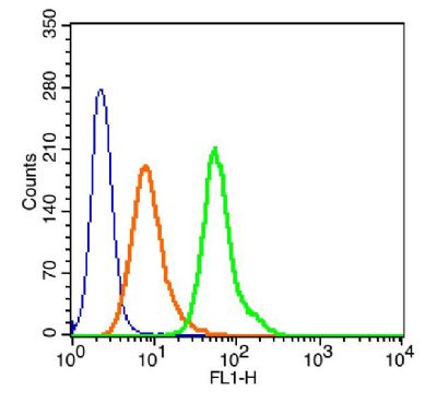
Blank control(blue): 293T(fixed with 2% paraformaldehyde (10 min) and then permeabilized with ice-cold 90% methanol for 30 min on ice).
Primary Antibody: Rabbit Anti- Phospho-c-Fos (Thr325)/AF488 Conjugated antibody (SL3154R /AF488), Dilution: 1μg in 100 μL 1X PBS containing 0.5% BSA;
Isotype Control Antibody: Rabbit IgG/FITC(orange) ,used under the same conditions.
|