[IF=2.952] Linyan Cheng. et al. Icariin attenuates thioacetamide‑induced bone loss via the RANKL‑p38/ERK‑NFAT signaling pathway. Mol Med Rep. 2022 Apr;25(4):1-11 WB ; Rat.
[IF=9.078] Tzu-Hsiang Lin. et al. A bilineage thermosensitive hydrogel system for stimulation of mesenchymal stem cell differentiation and enhancement of osteochondral regeneration. Compos Part B-Eng. 2022 Jan;:109614 ICC ; Rabbit.
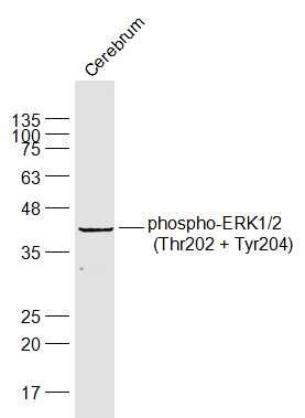
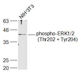
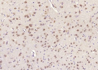
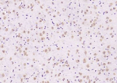
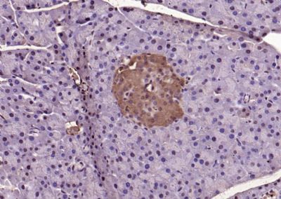
[IF=2.316] Song, Yue. et al. Seasonal expression of extracellular signal regulated kinases in the colon of wild ground squirrels (Spermophilus dauricus). Mol Biol Rep. 2022 Jan;:1-7 WB,IHC ; Squirrel.
[IF=5.279] Lei Wang. et al. Lactobacillus plantarum DP189 Reduces α-SYN Aggravation in MPTP-Induced Parkinson’s Disease Mice via Regulating Oxidative Damage, Inflammation, and Gut Microbiota Disorder. J Agr Food Chem. 2022;70(4):1163–1173 WB ; Mouse.
[IF=5.455] Zhang, Rongrong. et al. Compound traditional Chinese medicine dermatitis ointment ameliorates inflammatory responses and dysregulation of itch-related molecules in atopic dermatitis. Chin Med-Uk. 2022 Dec;17(1):1-19 WB ; Mouse.
[IF=5.31] Ning Zhong. et al. Apatinib inhibits the growth of small cell lung cancer via a mechanism mediated by VEGF, PI3K/Akt and Ki-67/CD31. 2021 Sep 30 IF ; mouse.
[IF=3.332] Zhixia Jia. et al. Baicalin Ameliorates Chronic Unpredictable Mild Stress-Induced Depression through the BDNF/ERK/CREB Signaling Pathway. Behav Brain Res. 2021 Jul;:113463 WB ; Mouse.
[IF=4.105] Ai Jin. et al. TSLP-induced collagen type-I synthesis through STAT3 and PRMT1 is sensitive to calcitriol in human lung fibroblasts. Bba-Mol Cell Res. 2021 Jun;:119083 WB ; Human.
[IF=4.225] Qihe Tang. et al. Bergenin Monohydrate Attenuates Inflammatory Response via MAPK and NF-κB Pathways Against Klebsiella pneumonia Infection. Front Pharmacol. 2021; 12: 651664 WB ; Mouse.
[IF=2.405] Keiichi Hiramoto. et al. Glycyrrhizin ameliorates melanoma cell extravasation into mouse lungs by regulating signal transduction through HMGB1 and its receptors. 2021 Mar 09 WB ; Mouse.
[IF=6.126] Xiaosu Chen. et al. DCBLD2 Mediates Epithelial-Mesenchymal Transition-Induced Metastasis by Cisplatin in Lung Adenocarcinoma. Cancers. 2021 Jan;13(6):1403 WB ; Human.
[IF=3.606] Xuejiao Zhou. et al. The novel ALK inhibitor ZX‐29 induces apoptosis through inhibiting ALK and inducing ROS‐mediated endoplasmic reticulum stress in Karpas299 cells. 2020 Nov 02 WB ; Human.
[IF=7.076] Yajing Lv. et al. Nucleotide de novo synthesis increases breast cancer stemness and metastasis via cGMP-PKG-MAPK signaling pathway. Plos Biol. 2020 Nov;18(11):e3000872 WB ; Mouse.
[IF=3.216] Chao Lu. et al. Inhibition of Pre-B Cell Colony Enhancing Factor Reduces Lung Injury in Rats Receiving Cardiopulmonary Bypass. Drug Des Dev Ther. 2021; 15: 51–60 WB ; Rat.
[IF=4.171] Gu F et al. Lactobacillus casei improves depression-like behavior in chronic unpredictable mild stress-induced rats by the BDNF-TrkB signal pathway and the intestinal microbiota. Food Funct. 2020 Jul 1;11(7):6148-6157. WB ; Rat.
[IF=2.571] Lijing Wanget al. 1-(4-((5-chloro-4-((2-(isopropylsulfonyl)phenyl)amino)pyrimidin-2-yl)amino)-3-methoxyphenyl)-3-(2-(dimethylamino)ethyl)imidazolidin-2-one (ZX-42), a novel ALK inhibitor, induces apoptosis and protective autophagy in H2228 cells. J Pharm Pharmacol
. 2020 Oct;72(10):1370-1382. WB ; Human.
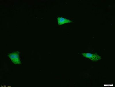
[IF=3.743] Liu F et al. Clinical and biological significances of heat shock protein 90 (Hsp90) in human nasopharyngeal carcinoma cells and anti-cancer effects of Hsp90 inhibitor. Biomed Pharmacother. 2019 Oct 18;120:109533. ICF ; Human.
[IF=3.266] Ma Q et al. Vitamin B5 inhibit RANKL induced osteoclastogenesis and ovariectomy induced osteoporosis by scavenging ROS generation. Am J Transl Res. 2019 Aug 15;11(8):5008-5018. eCollection 2019. WB ; Mouse.
[IF=2.394] Liu W et al. Synergistic effect of tolfenamic acid and glycyrrhizic acid on TPA-induced skin inflammation in mice. MedChemComm.2019. IHSLCP ; Mouse.
[IF=2.535] Lei J et al. LncRNA SNHG1 Alleviates IL-1β-induced Osteoarthritis by Inhibiting miR-16-5p-mediated p38 MAPK and NF-κB Signaling Pathways. Biosci Rep. 2019 Aug 5. pii: BSR20191523. WB ; Human.
[IF=3.118] Fu S et al. Berberine suppresses mast cell-mediated allergic responses via regulating FcɛRI-mediated and MAPK signaling.Int Immunopharmacol. 2019 Jun;71:1-6. WB ; Human.
[IF=2.342] Liu W et al. Glycyrrhizic acid from licorice down-regulates inflammatory responses via blocking MAPK and PI3K/Akt-dependent NF-κB signalling pathways in TPA-induced skin inflammation. MedChemComm. 2018. IHSLCP ; Mouse.
[IF=1.374] Zheng et al. Ginsenoside Rb1 improves cardiac function and remodeling in heart failure. (2017) Exp.Ani. 66:217-228 WB ; Rat.
[IF=5.008] Tan et al. ART3 regulates triple-negative breast cancer cell function via activation of Akt and ERK pathways. (2016) Oncotarge. 7:46589-46602 WB,IHC ; Human.
[IF=4.2] Zhang, Fengwei, et al. "Seasonal Expression of Oxytocin and Oxytocin Receptor in the Scented Gland of Male Muskrat (Ondatra zibethicus)." Scientific Reports 7.1 (2017): 16627. IHSLCP ; Others.
[IF=1.92] Liu, Chen, et al. "Pulmonary artery denervation improves pulmonary arterial hypertension induced right ventricular dysfunction by modulating the local renin-angiotensin-aldosterone system." BMC Cardiovascular Disorders 16.1 (2016): 192. WB ; Dog.
[IF=3.84] Geng, Shanshan, et al. "Medium-chain triglyceride ameliorates insulin resistance and inflammation in high fat diet-induced obese mice." European Journal of Nutrition (2015): 1-10. WB ; Mouse.
[IF=4.122] Jeon et al. Activation of the complement system in an osteosarcoma cell line promotes angiogenesis through enhanced production of growth factors. (2018) Sci.Rep. 8:5415 WB ;
[IF=2.656] Fang et al. Upregulation of long noncoding RNA CCAT1-L promotes epithelial-mesenchymal transition in gastric adenocarcinoma. (2018) Onco.Targets.Ther. 11:5647-5655 WB ; Human.
[IF=2.342] Liu W et al. Glycyrrhizic acid from licorice down-regulates inflammatory responses via blocking MAPK and PI3K/Akt-dependent NF-κB signalling pathways in TPA-induced skin inflammation. Medchemcomm. 2018 Jul 19;9(9):1502-1510. IHSLCP ; Mouse.
[IF=4.486] Jianye Yang. et al. Astragalus polysaccharide attenuates LPS-related inflammatory osteolysis by suppressing osteoclastogenesis by reducing the MAPK signalling pathway. 2021 Jun 02 WB ; Mouse.
[IF=5.279] Wei Liu. et al. Glycolysis and Reactive Oxygen Species Production Participate in T-2 Toxin-Stimulated Chicken Heterophil Extracellular Traps. J Agr Food Chem. 2021;XXXX(XXX):XXX-XXX WB ; Chicken.
[IF=4.75] Rosenzweig, Derek H., Sing J. Ou, and Thomas M. Quinn. ʺP38 mitogen activated protein kinase promotes dedifferentiation of primary articular chondrocytes in monolayer culture.ʺ Journal of Cellular and Molecular Medicine (2013). WB ; Bovine.
[IF=3.352] Zhikai Wu. et al. Fumonisin B1 induces chicken heterophil extracellular traps mediated by PAD4 enzyme and P2 × 1 receptor. Poultry Sci. 2021 Oct;:101550 WB ; chicken .
[IF=3.738] Lili Liu. et al. Rutin Ameliorates Cadmium-Induced Necroptosis in the Chicken Liver via Inhibiting Oxidative Stress and MAPK/NF-κB Pathway. 2021 Jun 06 WB ; Chicken.
[IF=3.943] Xiuhong Wang. et al. Protective effect of combination of anakinra and MCC950 against acute lung injury is achieved through suppression of the NF-κB-mediated-MAPK and NLRP3-caspase pathways. Int Immunopharmacol. 2021 Aug;97:107506 WB ; Mouse.
[IF=1.918] Jingwen Zhou. et al. Zi Qi Decoction Alleviates Liver Fibrosis by Inhibiting the Toll-Like Receptor 4 (TLR4)-Related Nuclear Factor kappa b (NF-κB) and Mitogen-Activated Protein Kinase (MAPK) Signaling Pathways. Med Sci Monitor. 2021; 27: e929438-1–e929438-12 WB ; Rat.
[IF=3.641] Wenbo Ge. et al. 17β-estradiol protects sheep oviduct epithelial cells against lipopolysaccharide-induced inflammation in vitro. Mol Immunol. 2020 Nov;127:21 WB ; Sheep.
[IF=14.528] Dejun Xu. et al. Melatonin protects mouse testes from palmitic acid‐induced lipotoxicity by attenuating oxidative stress and DNA damage in a SIRT1‐dependent manner. J Pineal Res. 2020 Nov;69(4):e12690 WB ; Mouse.
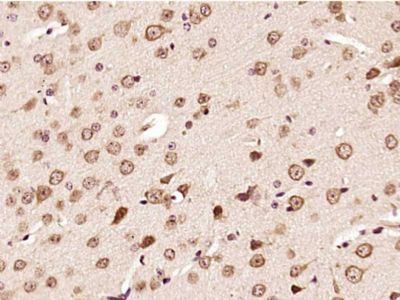
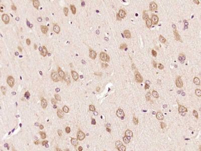
[IF=8.109] Li X et al. Disseminated Melanoma Cells Transdifferentiate into Endothelial Cells in Intravascular Niches at Metastatic Sites. Cell Rep. 2020 Jun 16;31(11):107765. IHF ; Mouse.
[IF=4.171] Gu F et al. Lactobacillus casei improves depression-like behavior in chronic unpredictable mild stress-induced rats by BDNF-TrkB signal pathway and the intestinal microbiota. Food Funct
. 2020 Jul 1;11(7):6148-6157. WB ; Rat.
[IF=2.445] Yuan Z et al. Seasonal expressions of oxytocin and oxytocin receptor in the epididymides in the wild ground squirrels (Citellus Dauricus Brandt). Gen Comp Endocrinol. 2020 Apr 1;289:113391. WB&IHSLCP ; Wild ground squirrel.
[IF=4.658] Zhang J et al. Down‐regulation of Suv39h1 attenuates neointima formation after carotid artery injury in diabetic rats. J Cell Mol Med. 2019 Nov 17. WB ; Rat.
[IF=2.53] Li Y et al. Ecdysterone Accelerates Healing of Radiation-Induced Oral Mucositis in Rats by Increasing Matrix Cell Proliferation. Radiat Res. 2019 Mar;191(3):237-244. WB ; Rat.
[IF=2.332] Zhao X et al. Total flavones of fermentation broth by co-culture of Coprinus comatus and Morchella esculenta induces an anti-inflammatory effect on LPS-stimulated RAW264.7 macrophages cells via the MAPK signaling pathway.(2018) Microb Pathog.125:431-437. WB ; Mouse.
[IF=1.77] Xing et al. Increased expression of receptor for advanced glycation end-products worsens focal brain ischemia in diabetic rats. (2012) Neural.Regen.Res. 7:1000-5 WB ; Rat.
[IF=0.764] Chen et al. Downregulation of MACC1 expression enhances cisplatin sensitivity in SKOSLV3/DDP cells. (2015) Genet.Mol.Re. 14:17134-44 WB ; Human.
[IF=2.62] Liu, Lu, et al. "Anthraquinone derivative exerted hormetic effect on the apoptosis in oxygen-glucose deprivation-induced PC12 cells via ERK and Akt activated Nrf2/HO-1 signaling pathway." Chemico-Biological Interactions 262 (2017): 1-11. WB ; Rat.
[IF=1.43] Wang, Xiao-yan, et al. "AMD620 attenuates MMP-3 and MMP-9 expressions and prevents cartilage degradation in a monosodium iodoacetate-induced rat model of temporomandibular osteoarthritis." Journal of Oral and Maxillofacial Surgery (2016). IHSLCP ; Rat.
[IF=3.04] Chen, Wei, et al. "Mycobacterium tuberculosis PE25/PPE41 protein complex induces activation and maturation of dendritic cells and drives Th2-biased immune responses." Medical Microbiology and Immunology (2015): 1-13. WB ; Mouse.