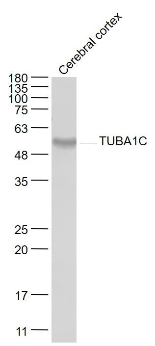
Sample:
Cerebral cortex (Mouse) Lysate at 40 ug
Primary: Anti- TUBA1C (SL5118R) at 1/1000 dilution
Secondary: IRDye800CW Goat Anti-Rabbit IgG at 1/20000 dilution
Predicted band size: 49 kD
Observed band size: 52 kD
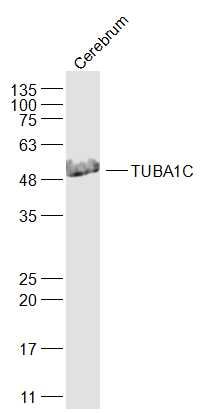
Sample:
Cerebrum (Mouse) Lysate at 40 ug
Primary: Anti-TUBA1C (SL5118R) at 1/1000 dilution
Secondary: IRDye800CW Goat Anti-Rabbit IgG at 1/20000 dilution
Predicted band size: 49 kD
Observed band size: 49 kD
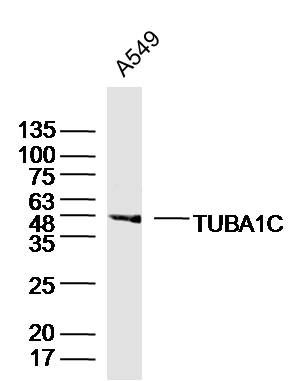
Sample:
A549 Cell (Human) Lysate at 30 ug
Primary: Anti- TUBA1C (SL5118R)at 1/300 dilution
Secondary: IRDye800CW Goat Anti-Rabbit IgG at 1/20000 dilution
Predicted band size: 49kD
Observed band size: 49kD
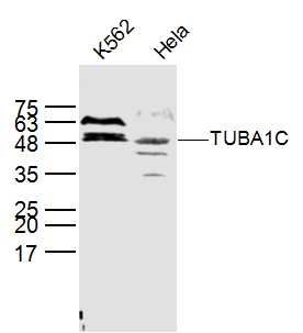
Sample:
K562(Human) Cell Lysate at 40 ug
Hela(Human) Cell Lysate at 40 ug
Primary: Anti-TUBA1C (SL5118R) at 1/300 dilution
Secondary: IRDye800CW Goat Anti-Rabbit IgG at 1/20000 dilution
Predicted band size: 49 kD
Observed band size: 49 kD
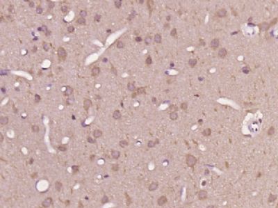
Paraformaldehyde-fixed, paraffin embedded (rat brain tissue); Antigen retrieval by boiling in sodium citrate buffer (pH6.0) for 15min; Block endogenous peroxidase by 3% hydrogen peroxide for 20 minutes; Blocking buffer (normal goat serum) at 37°C for 30min; Antibody incubation with (TUBA1C) Polyclonal Antibody, Unconjugated (SL5118R) at 1:400 overnight at 4°C, followed by operating according to SP Kit(Rabbit) (sp-0023) instructionsand DAB staining.
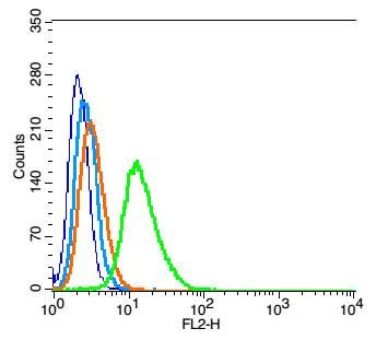
Blank control(blue): RSC96 (fixed with 2% paraformaldehyde (10 min),then permeabilized with 90% ice-cold methanol for 30 min on ice).
Primary Antibody:Rabbit Anti- TUBA1C antibody(SL5118R), Dilution: 1μg in 100 μL 1X PBS containing 0.5% BSA;
Isotype Control Antibody: Rabbit IgG(orange) ,used under the same conditions );
Secondary Antibody: Goat anti-rabbit IgG-PE(white blue), Dilution: 1:200 in 1 X PBS containing 0.5% BSA.
|