AIM2 is a member of the IFI20X /IFI16 family. It plays a putative role in tumorigenic reversion and may control cell proliferation. Interferon-gamma induces expression of AIM2.
Function:
Involved in innate immune response by recognizing cytosolic double-stranded DNA and inducing caspase-1-activating inflammasome formation in macrophages. Upon binding to DNA is thought to undergo oligomerization and to associate with PYCARD initiating the recruitment of caspase-1 precusrsor and processing of interleukin-1 beta and interleukin-18. Detects cytosolic dsDNA of viral and bacterial origin in a non-sequence-specific manner. Can also trigger PYCARD-dependent, caspase-1-independent cell death that involves caspase-8.
Subunit:
Self-associates; forms homooligomers in response to cytosolic dsDNA and the dsDNA seems to serve as oligomerization platform. Component of the AIM2 inflammasome complex. Interacts with PYCARD. Interacts with IFI16 (By similarity). Interacts with EIF2AK2/PKR (By similarity).
Subcellular Location:
Nucleus (By similarity). Cytoplasm.
Tissue Specificity:
Expressed in spleen, small intestine, peripheral blood leukocytes, and testis.
Similarity:
Belongs to the HIN-200 family.
Contains 1 DAPIN domain.
Contains 1 HIN-200 domain.
SWISS:
Q91VJ1
Gene ID:
383619
Database links:
Entrez Gene: 9447 Human
Entrez Gene: 383619 Mouse
Entrez Gene: 304987 Rat
Omim: 604578 Human
SwissProt: O14862 Human
SwissProt: Q91VJ1 Mouse
Unigene: 281898 Human
Unigene: 733411 Human
Unigene: 131453 Mouse
| Picture |
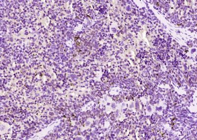
Paraformaldehyde-fixed, paraffin embedded (mouse spleen); Antigen retrieval by boiling in sodium citrate buffer (pH6.0) for 15min; Block endogenous peroxidase by 3% hydrogen peroxide for 20 minutes; Blocking buffer (normal goat serum) at 37°C for 30min; Antibody incubation with (AIM2) Polyclonal Antibody, Unconjugated (SL5986R) at 1:200 overnight at 4°C, followed by operating according to SP Kit(Rabbit) (sp-0023) instructionsand DAB staining.
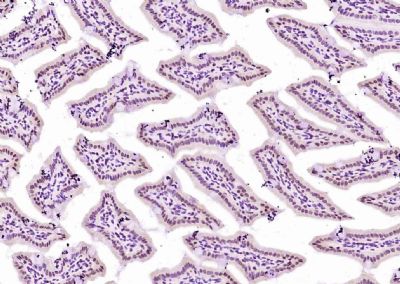
Paraformaldehyde-fixed, paraffin embedded (mouse intestine); Antigen retrieval by boiling in sodium citrate buffer (pH6.0) for 15min; Block endogenous peroxidase by 3% hydrogen peroxide for 20 minutes; Blocking buffer (normal goat serum) at 37°C for 30min; Antibody incubation with (AIM2) Polyclonal Antibody, Unconjugated (SL5986R) at 1:200 overnight at 4°C, followed by operating according to SP Kit(Rabbit) (sp-0023) instructionsand DAB staining.
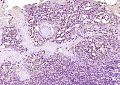
Paraformaldehyde-fixed, paraffin embedded (human tonsil); Antigen retrieval by boiling in sodium citrate buffer (pH6.0) for 15min; Block endogenous peroxidase by 3% hydrogen peroxide for 20 minutes; Blocking buffer (normal goat serum) at 37°C for 30min; Antibody incubation with (AIM2) Polyclonal Antibody, Unconjugated (SL5986R) at 1:200 overnight at 4°C, followed by operating according to SP Kit(Rabbit) (sp-0023) instructionsand DAB staining.
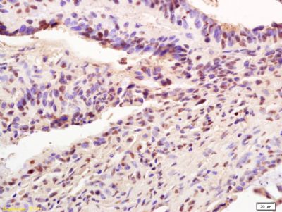
Tissue/cell: human colon carcinoma; 4% Paraformaldehyde-fixed and paraffin-embedded;
Antigen retrieval: citrate buffer ( 0.01M, pH 6.0 ), Boiling bathing for 15min; Block endogenous peroxidase by 3% Hydrogen peroxide for 30min; Blocking buffer (normal goat serum,SLC0005) at 37℃ for 20 min;
Incubation: Anti-AIM2 Polyclonal Antibody, Unconjugated(SL5986R) 1:200, overnight at 4°C, followed by conjugation to the secondary antibody(SP-0023) and DAB(SLC0010) staining
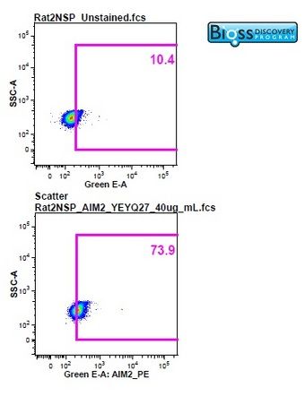
Image was kindly submitted by a researcher at Duke University Medical Center. Rat splenocytes stained with Rabbit Anti-AIM2 Polyclonal Antibody, PE conjugated (SL5986R-PE)at 1:25.
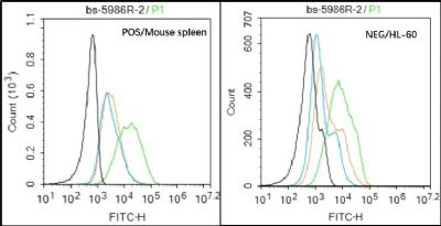
Black line : Positive blank control (Mouse spleen); Negative blank control (HL60)
Green line : Primary Antibody (Rabbit Anti-AIM2 antibody (SL5986R) )
Orange line:Isotype Control Antibody (Rabbit IgG) .
Blue line : Secondary Antibody (Goat anti-rabbit IgG-AF488)
Mouse spleen(Positive)and HL60(Negative control)cells (black) were fixed with 4% PFA for 10min at room temperature, permeabilized with PBST for 20 min at room temperature, and incubated in 5% BSA blocking buffer for 30 min at room temperature. Cells were then stained with AIM2 Antibody(SL5986R)at 1:50 dilution in blocking buffer and incubated for 30 min at room temperature, washed twice with 2% BSA in PBS, followed by secondary antibody(blue) incubation for 40 min at room temperature. Acquisitions of 20,000 events were performed. Cells stained with primary antibody (green), and isotype control (orange).
|
|
|