Home > Product > Antibody > Rabbit Anti-TCF3 antibody
TFE2_HUMAN; Transcription factor E2-alpha; BHLHB21; E2A; ITF1; E47; p75; AGM8; VDIR; AGM8A; AGM8B; TCF-3; bHLHb21Class B basic helix-loop-helix protein 21 (bHLHb21); Immunoglobulin enhancer-binding factor E12/E47; Immunoglobulin transcription factor 1;
Cat:
SL6065R
Species Reactivity:
Human,Mouse,Rat,(predicted: Dog,Cow,Horse,)
Immunogen:
KLH conjugated synthetic peptide derived from human TCF3/E2A:301-400/654
Format:
Liquid
Storage instructions:
Shipped at 4℃. Store at -20 °C for one year. Avoid repeated freeze/thaw cycles.
Concentration:
1mg/ml
Clonality:
Polyclonal
Isotype:
IgG
Applications:
WB=1:500-2000ELISA=1:5000-10000IHC-P=1:100-500IHC-F=1:100-500IF=1:100-500(Paraffin sections need to do antigen repair)not yet tested in other applications.optimal dilutions/concentrations should be determined by the end user.
Host:
Rabbit
Product Overview:
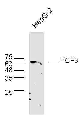
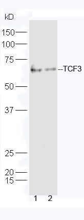
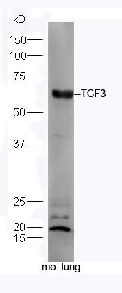
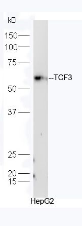
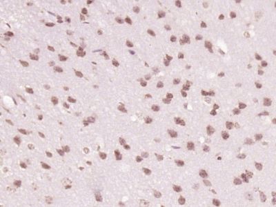
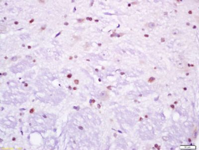
Sample: HepG2 Cell (Human) Lysate at 40 ugPrimary: Anti-TCF3 (SL6065R) at 1/300 dilutionSecondary: IRDye800CW Goat Anti-Rabbit IgG at 1/20000 dilutionPredicted band size: 67 kDObserved band size: 67 kDSample: A549 Cell (Human) Lysate at 40 ugRaji Cell (Human) Lysate at 40 ugPrimary: Anti-TCF3 (SL6065R) at 1/300 dilutionSecondary: HRP conjugated Goat-Anti-rabbit IgG (SL0295G-HRP) at 1/5000 dilutionPredicted band size: 67 kDObserved band size: 67 kDSample: Lung (Mouse) Lysate at 40 ugPrimary: Anti-TCF3 (SL6065R) at 1/300 dilutionSecondary: HRP conjugated Goat-Anti-rabbit IgG (SL0295G-HRP) at 1/5000 dilutionPredicted band size: 67 kDObserved band size: 67 kDSample: HepG2 Cell (Human) Lysate at 40 ugPrimary: Anti-TCF3 (SL6065R) at 1/300 dilutionSecondary: HRP conjugated Goat-Anti-rabbit IgG (SL0295G-HRP) at 1/5000 dilutionPredicted band size: 67 kDObserved band size: 67 kDParaformaldehyde-fixed, paraffin embedded (Mouse brain); Antigen retrieval by boiling in sodium citrate buffer (pH6.0) for 15min; Block endogenous peroxidase by 3% hydrogen peroxide for 20 minutes; Blocking buffer (normal goat serum) at 37°C for 30min; Antibody incubation with (TCF3) Polyclonal Antibody, Unconjugated (SL6065R) at 1:400 overnight at 4°C, followed by operating according to SP Kit(Rabbit) (sp-0023) instructionsand DAB staining.Tissue/cell: rat brain tissue; 4% Paraformaldehyde-fixed and paraffin-embedded; Antigen retrieval: citrate buffer ( 0.01M, pH 6.0 ), Boiling bathing for 15min; Block endogenous peroxidase by 3% Hydrogen peroxide for 30min; Blocking buffer (normal goat serum,SLC0005) at 37℃ for 20 min; Incubation: Anti-TCF3 Polyclonal Antibody, Unconjugated(SL6065R) 1:200, overnight at 4°C, followed by conjugation to the secondary antibody(SP-0023) and DAB(SLC0010) staining
More
Unit:
Price: $
Product PDFs
Datasheet:


This gene encodes a member of the E protein (class I) family of helix-loop-helix transcription factors. E proteins activate transcription by binding to regulatory E-box sequences on target genes as heterodimers or homodimers, and are inhibited by heterodimerization with inhibitor of DNA-binding (class IV) helix-loop-helix proteins. E proteins play a critical role in lymphopoiesis, and the encoded protein is required for B and T lymphocyte development. Deletion of this gene or diminished activity of the encoded protein may play a role in lymphoid malignancies. This gene is also involved in several chromosomal translocations that are associated with lymphoid malignancies including pre-B-cell acute lymphoblastic leukemia (t(1;19), with PBX1), childhood leukemia (t(19;19), with TFPT) and acute leukemia (t(12;19), with ZNF384). Alternatively spliced transcript variants encoding multiple isoforms have been observed for this gene, and a pseudogene of this gene is located on the short arm of chromosome 9. [provided by RefSeq, Sep 2011]

Function:
Transcriptional regulator. Involved in the initiation of neuronal differentiation. Heterodimers between TCF3 and tissue-specific basic helix-loop-helix (bHLH) proteins play major roles in determining tissue-specific cell fate during embryogenesis, like muscle or early B-cell differentiation. Dimers bind DNA on E-box motifs: 5'-CANNTG-3'. Binds to the kappa-E2 site in the kappa immunoglobulin gene enhancer. Binds to IEB1 and IEB2, which are short DNA sequences in the insulin gene transcription control region.

Subunit:
Forms a heterodimer with ASH1 and TWIST2. Isoform E12 interacts with RALGAPA1 and FIGLA. Efficient DNA binding requires dimerization with another bHLH protein. Component of a nuclear TAL-1 complex composed at least of CSFA2T3, LDB1, TAL1 and TCF3. Interacts with NEUROD2, PTF1A and TGFB1I1 (By similarity). Homodimer. Heterodimer. Forms a heterodimer with NEUROD1; the heterodimer is inhibited in presence of ID2, but not NR0B2, to E-box element. Interacts with EP300 and UBE2I.

Subcellular Location:
Nucleus.

Post-translational modifications:
Phosphorylated following NGF stimulation (By similarity).

DISEASE:
Note=Chromosomal aberrations involving TCF3 are cause of forms of pre-B-cell acute lymphoblastic leukemia (B-ALL). Translocation t(1;19)(q23;p13.3) with PBX1. TCF3-PBX1 transforms cells by constitutively activating transcription of genes regulated by PBX1 or by other members of the PBX protein family. Translocation t(17;19)(q22;p13.3) with HLF. Inversion inv(19)(p13;q13) with TFPT.

Similarity:
Contains 1 bHLH (basic helix-loop-helix) domain.

SWISS:
P15923

Gene ID:
6929

Database links:

Entrez Gene: 6929 Human

Entrez Gene: 21423 Mouse

Entrez Gene: 171046 Rat

SwissProt: P15923 Human

SwissProt: P3166 Mouse

SwissProt: P21677 Rat


Picture

Sample: HepG2 Cell (Human) Lysate at 40 ug
Primary: Anti-TCF3 (SL6065R) at 1/300 dilution
Secondary: IRDye800CW Goat Anti-Rabbit IgG at 1/20000 dilution
Predicted band size: 67 kD
Observed band size: 67 kD
Sample:
A549 Cell (Human) Lysate at 40 ug
Raji Cell (Human) Lysate at 40 ug
Primary: Anti-TCF3 (SL6065R) at 1/300 dilution
Secondary: HRP conjugated Goat-Anti-rabbit IgG (SL0295G-HRP) at 1/5000 dilution
Predicted band size: 67 kD
Observed band size: 67 kD
Sample: Lung (Mouse) Lysate at 40 ug
Primary: Anti-TCF3 (SL6065R) at 1/300 dilution
Secondary: HRP conjugated Goat-Anti-rabbit IgG (SL0295G-HRP) at 1/5000 dilution
Predicted band size: 67 kD
Observed band size: 67 kD
Sample: HepG2 Cell (Human) Lysate at 40 ug
Primary: Anti-TCF3 (SL6065R) at 1/300 dilution
Secondary: HRP conjugated Goat-Anti-rabbit IgG (SL0295G-HRP) at 1/5000 dilution
Predicted band size: 67 kD
Observed band size: 67 kD
Paraformaldehyde-fixed, paraffin embedded (Mouse brain); Antigen retrieval by boiling in sodium citrate buffer (pH6.0) for 15min; Block endogenous peroxidase by 3% hydrogen peroxide for 20 minutes; Blocking buffer (normal goat serum) at 37°C for 30min; Antibody incubation with (TCF3) Polyclonal Antibody, Unconjugated (SL6065R) at 1:400 overnight at 4°C, followed by operating according to SP Kit(Rabbit) (sp-0023) instructionsand DAB staining.
Tissue/cell: rat brain tissue; 4% Paraformaldehyde-fixed and paraffin-embedded;
Antigen retrieval: citrate buffer ( 0.01M, pH 6.0 ), Boiling bathing for 15min; Block endogenous peroxidase by 3% Hydrogen peroxide for 30min; Blocking buffer (normal goat serum,SLC0005) at 37℃ for 20 min;
Incubation: Anti-TCF3 Polyclonal Antibody, Unconjugated(SL6065R) 1:200, overnight at 4°C, followed by conjugation to the secondary antibody(SP-0023) and DAB(SLC0010) staining
Product Feedback Wall
Message :
Your Email :
Copyright © 2007-2018 Sunlong Medical All Rights Reserved.