Home > Product > Antibody > Rabbit Anti-JAMC antibody
CAM; JAM 2; JAM 3; JAM C; JAM-2; JAM-3; JAM-C; JAM2; Jam3; JAM3_HUMAN; JAMC; Junction adhesion molecule C; Junctional adhesion molecule 3; Junctional adhesion molecule 3 precursor; Junctional adhesion molecule C.
Cat:
SL11086R
Species Reactivity:
Human,Mouse,Rat,(predicted: Dog,Pig,Cow,Horse,)
Immunogen:
KLH conjugated synthetic peptide derived from human JAMC:31-130/310<Extracellular>
Format:
Liquid
Storage instructions:
Shipped at 4℃. Store at -20 °C for one year. Avoid repeated freeze/thaw cycles.
Concentration:
1mg/ml
Clonality:
Polyclonal
Isotype:
IgG
Applications:
WB=1:500-2000ELISA=1:5000-10000IHC-P=1:100-500IHC-F=1:100-500ICC=1:100-500IF=1:100-500(Paraffin sections need to do antigen repair)not yet tested in other applications.optimal dilutions/concentrations should be determined by the end user.
Host:
Rabbit
Product Overview:
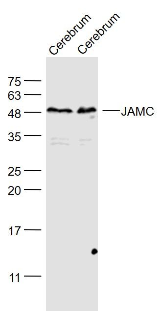
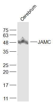
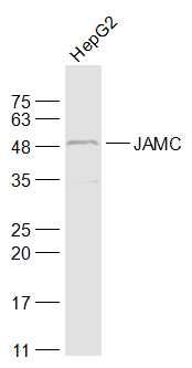
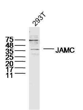
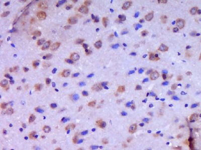
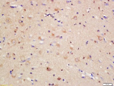
Sample: Cerebrum (Mouse) Lysate at 40 ugCerebrum (Rat) Lysate at 40 ugPrimary: Anti- JAMC (SL11086R) at 1/1000 dilutionSecondary: IRDye800CW Goat Anti-Rabbit IgG at 1/20000 dilutionPredicted band size: 31 kDObserved band size: 49 kDSample: Cerebrum (Mouse) Lysate at 40 ugPrimary: Anti-JAMC (SL11086R) at 1/1000 dilutionSecondary: IRDye800CW Goat Anti-Rabbit IgG at 1/20000 dilutionPredicted band size: 31 kDObserved band size: 49 kDSample: HepG2(Human) Cell Lysate at 30 ugPrimary: Anti-JAMC (SL11086R) at 1/1000 dilutionSecondary: IRDye800CW Goat Anti-Rabbit IgG at 1/20000 dilutionPredicted band size: 31 kDObserved band size: 49 kDSample: 293T Cell (Human) Lysate at 40 ugPrimary: Anti-JAMC (SL11086R) at 1/300 dilutionSecondary: IRDye800CW Goat Anti-Rabbit IgG at 1/20000 dilutionPredicted band size: 31 kDObserved band size: 36 kDParaformaldehyde-fixed, paraffin embedded (Mouse brain); Antigen retrieval by boiling in sodium citrate buffer (pH6.0) for 15min; Block endogenous peroxidase by 3% hydrogen peroxide for 20 minutes; Blocking buffer (normal goat serum) at 37°C for 30min; Antibody incubation with (JAMC) Polyclonal Antibody, Unconjugated (SL11086R) at 1:400 overnight at 4°C, followed by operating according to SP Kit(Rabbit) (sp-0023) instructionsand DAB staining.Tissue/cell: rat brain tissue; 4% Paraformaldehyde-fixed and paraffin-embedded; Antigen retrieval: citrate buffer ( 0.01M, pH 6.0 ), Boiling bathing for 15min; Block endogenous peroxidase by 3% Hydrogen peroxide for 30min; Blocking buffer (normal goat serum,SLC0005) at 37℃ for 20 min; Incubation: Anti-JAMC Polyclonal Antibody, Unconjugated(SL11086R) 1:200, overnight at 4°C, followed by conjugation to the secondary antibody(SP-0023) and DAB(SLC0010) staining
More
Unit:
Price: $
Product PDFs
Datasheet:


Tight junctions represent one mode of cell-to-cell adhesion in epithelial or endothelial cell sheets, forming continuous seals around cells and serving as a physical barrier to prevent solutes and water from passing freely through the paracellular space. The protein encoded by this immunoglobulin superfamily gene member is localized in the tight junctions between high endothelial cells. Unlike other proteins in this family, the this protein is unable to adhere to leukocyte cell lines and only forms weak homotypic interactions. The encoded protein is a member of the junctional adhesion molecule protein family and acts as a receptor for another member of this family. A mutation in an intron of this gene is associated with hemorrhagic destruction of the brain, subependymal calcification, and congenital cataracts. Alternative splicing results in multiple transcript variants.[provided by RefSeq, Apr 2011].

Function:
Participates in cell-cell adhesion. It is a counter-receptor for ITGAM, mediating leukocyte-platelet interactions and is involved in the regulation of transepithelial migration of polymorphonuclear neutrophils (PMN). The soluble form is a mediator of angiogenesis.

Subunit:
Interacts with JAM2. Interacts with ITGAM.

Subcellular Location:
Cell membrane; Single-pass type I membrane protein (Potential). Cell junction, desmosome. Secreted, extracellular space. Note=In epithelial cells, it is expressed at desmosomes but not at tight junctions. Localizes at the cell surface of endothelial cells; treatment of endothelial cells with vascular endothelial growth factor stimulates recruitment of JAM3 to cell-cell contacts.

Tissue Specificity:
Highest expression in placenta, brain and kidney. Significant expression is detected on platelets. Expressed in intestinal mucosa cells. Expressed in the vascular endothelium. Found in serum (at protein level). Also detected in the synovial fluid of patients with rheumatoid arthritis, psoriatic arthritis or ostearthritis (at protein level).

Post-translational modifications:
Proteolytically cleaved from endothelial cells surface into a soluble form by ADAM10 and ADAM17; the release of soluble JAM3 is increased by proinflammatory factors.

DISEASE:
Defects in JAM3 are the cause of hemorrhagic destruction of the brain with subependymal calcification and cataracts (HDBSCC) [MIM:613730]. A syndrome characterized by congenital cataracts and severe brain abnormalities apparently resulting from hemorrhagic destruction of the brain tissue, including the cerebral white matter and basal ganglia. Patients manifest profound developmental delay, and other neurologic features included seizures, spasticity, and hyperreflexia. Brain imaging shows multifocal intraparenchymal hemorrhage with associated liquefaction and massive cystic degeneration, and calcification in the subependymal region and in brain tissue.

Similarity:
Belongs to the immunoglobulin superfamily.
Contains 1 Ig-like C2-type (immunoglobulin-like) domain.
Contains 1 Ig-like SLVtype (immunoglobulin-like) domain.


SWISS:
Q9BX67

Gene ID:
83700

Database links:

Entrez Gene: 83700 Human

Entrez Gene: 83964 Mouse

Entrez Gene: 315509 Rat

Omim: 606871 Human

SwissProt: Q9BX67 Human

SwissProt: Q9D8B7 Mouse

SwissProt: Q68FQ2 Rat

Unigene: 150718 Human

Unigene: 728339 Human

Unigene: 28770 Mouse

Unigene: 104684 Rat


Picture

Sample:
Cerebrum (Mouse) Lysate at 40 ug
Cerebrum (Rat) Lysate at 40 ug
Primary: Anti- JAMC (SL11086R) at 1/1000 dilution
Secondary: IRDye800CW Goat Anti-Rabbit IgG at 1/20000 dilution
Predicted band size: 31 kD
Observed band size: 49 kD
Sample:
Cerebrum (Mouse) Lysate at 40 ug
Primary: Anti-JAMC (SL11086R) at 1/1000 dilution
Secondary: IRDye800CW Goat Anti-Rabbit IgG at 1/20000 dilution
Predicted band size: 31 kD
Observed band size: 49 kD
Sample:
HepG2(Human) Cell Lysate at 30 ug
Primary: Anti-JAMC (SL11086R) at 1/1000 dilution
Secondary: IRDye800CW Goat Anti-Rabbit IgG at 1/20000 dilution
Predicted band size: 31 kD
Observed band size: 49 kD
Sample: 293T Cell (Human) Lysate at 40 ug
Primary: Anti-JAMC (SL11086R) at 1/300 dilution
Secondary: IRDye800CW Goat Anti-Rabbit IgG at 1/20000 dilution
Predicted band size: 31 kD
Observed band size: 36 kD
Paraformaldehyde-fixed, paraffin embedded (Mouse brain); Antigen retrieval by boiling in sodium citrate buffer (pH6.0) for 15min; Block endogenous peroxidase by 3% hydrogen peroxide for 20 minutes; Blocking buffer (normal goat serum) at 37°C for 30min; Antibody incubation with (JAMC) Polyclonal Antibody, Unconjugated (SL11086R) at 1:400 overnight at 4°C, followed by operating according to SP Kit(Rabbit) (sp-0023) instructionsand DAB staining.
Tissue/cell: rat brain tissue; 4% Paraformaldehyde-fixed and paraffin-embedded;
Antigen retrieval: citrate buffer ( 0.01M, pH 6.0 ), Boiling bathing for 15min; Block endogenous peroxidase by 3% Hydrogen peroxide for 30min; Blocking buffer (normal goat serum,SLC0005) at 37℃ for 20 min;
Incubation: Anti-JAMC Polyclonal Antibody, Unconjugated(SL11086R) 1:200, overnight at 4°C, followed by conjugation to the secondary antibody(SP-0023) and DAB(SLC0010) staining
Product Feedback Wall
Message :
Your Email :
Copyright © 2007-2018 Sunlong Medical All Rights Reserved.