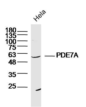
Sample: Hela Cell (Human) Lysate at 30 ug
Primary: Anti-PDE7A (SL11575R) at 1/300 dilution
Secondary: IRDye800CW Goat Anti-Rabbit IgG at 1/20000 dilution
Predicted band size: 55kD
Observed band size: 55kD
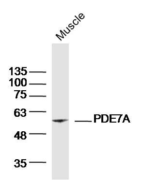
Sample: Muscle (Mouse)Lysate at 40 ug
Primary: Anti-PDE7A(SL11575R)at 1/300 dilution
Secondary: IRDye800CW Goat Anti-RabbitIgG at 1/20000 dilution
Predicted band size: 55kD
Observed band size: 55kD
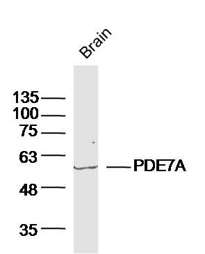
Sample:Brain (Mouse)Lysate at 40 ug
Primary: Anti-PDE7A(SL11575R)at 1/300 dilution
Secondary: IRDye800CW Goat Anti-RabbitIgG at 1/20000 dilution
Predicted band size: 55kD
Observed band size: 55kD
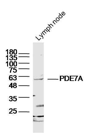
Sample: Lymph node (Mouse) Lysate at 40 ug
Primary: Anti-PDE7A (SL11575R) at 1/300 dilution
Secondary: IRDye800CW Goat Anti-Rabbit IgG at 1/20000 dilution
Predicted band size: 55kD
Observed band size: 55kD
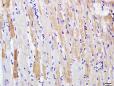
Paraformaldehyde-fixed, paraffin embedded (rat heart); Antigen retrieval by boiling in sodium citrate buffer (pH6.0) for 15min; Block endogenous peroxidase by 3% hydrogen peroxide for 20 minutes; Blocking buffer (normal goat serum) at 37°C for 30min; Antibody incubation with (PDE7A) Polyclonal Antibody, Unconjugated (SL11575R) at 1:500 overnight at 4°C, followed by a conjugated secondary (sp-0023) for 20 minutes and DAB staining.
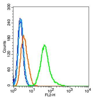
Blank control(blue): RSC96 cells(fixed with 2% paraformaldehyde (10 min) , then permeabilized with 90% ice-cold methanol for 30 min on ice).
Primary Antibody:Rabbit Anti- PDE7A antibody(SL11575R), Dilution: 1μg in 100 μL 1X PBS containing 0.5% BSA;
Isotype Control Antibody: Rabbit IgG(orange) ,used under the same conditions );
Secondary Antibody: Goat anti-rabbit IgG-PE(white blue), Dilution: 1:200 in 1 X PBS containing 0.5% BSA.
|