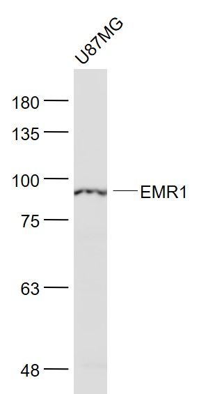
Sample:
U87MG(Human) Cell Lysate at 30 ug
Primary: Anti- EMR1 (SL7058R) at 1/1000 dilution
Secondary: IRDye800CW Goat Anti-Rabbit IgG at 1/20000 dilution
Predicted band size: 95 kD
Observed band size: 95 kD
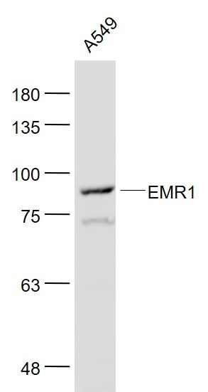
Sample:
A549(Human) Cell Lysate at 30 ug
Primary: Anti- EMR1 (SL7058R) at 1/1000 dilution
Secondary: IRDye800CW Goat Anti-Rabbit IgG at 1/20000 dilution
Predicted band size: 95 kD
Observed band size: 95 kD
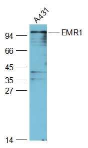
Sample:
A431(Human) Cell Lysate at 30 ug
Primary: Anti-EMR1 (SL7058R) at 1/2000 dilution
Secondary: IRDye800CW Goat Anti-Rabbit IgG at 1/20000 dilution
Predicted band size: 95 kD
Observed band size: 95 kD
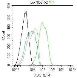
Blank control:A431.
Primary Antibody (green line): Rabbit Anti-ADGRE1 antibody (SL7058R)
Dilution: 2ug/Test;
Secondary Antibody : Goat anti-rabbit IgG-AF488
Dilution: 0.5ug/Test.
Protocol
The cells were incubated in 5%BSA to block non-specific protein-protein interactions for 30 min at room temperature .Cells stained with Primary Antibody for 30 min at room temperature. The secondary antibody used for 40 min at room temperature. Acquisition of 20,000 events was performed.
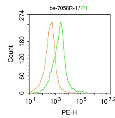
Blank control: Mouse kidney.
Primary Antibody (green line): Rabbit Anti-EMR1 antibody (SL7058R)
Dilution: 1μg /10^6 cells;
Isotype Control Antibody (orange line): Rabbit IgG .
Secondary Antibody : Goat anti-rabbit IgG-PE
Dilution: 1μg /test.
Protocol
The cells were incubated in 5%BSA to block non-specific protein-protein interactions for 30 min at at room temperature .Cells stained with Primary Antibody for 30 min at room temperature. The secondary antibody used for 40 min at room temperature. Acquisition of 20,000 events was performed.
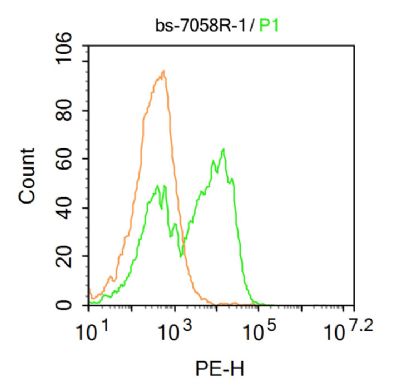
Blank control: Mouse brain.
Primary Antibody (green line): Rabbit Anti-EMR1 antibody (SL7058R)
Dilution: 1μg /10^6 cells;
Isotype Control Antibody (orange line): Rabbit IgG .
Secondary Antibody : Goat anti-rabbit IgG-PE
Dilution: 1μg /test.
Protocol
The cells were fixed with 4% PFA (10min at room temperature)and then permeabilized with 90% ice-cold methanol for 20 min at-20℃. The cells were then incubated in 5%BSA to block non-specific protein-protein interactions for 30 min at at room temperature .Cells stained with Primary Antibody for 30 min at room temperature. The secondary antibody used for 40 min at room temperature. Acquisition of 20,000 events was performed.
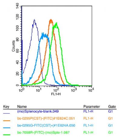
Positive control: mouse Splenocytes(2% Paraformaldehyde-fixed )
Isotype Control Antibody: Rabbit IgG
Dilution: 1μg in 100 μl 1X PBS containing 0.5% BSA;
Secondary Antibody: Goat anti-rabbit IgG-FITC;
Dilution: 1:200 in 1 X PBS containing 0.5% BSA;
Primary Antibody : rabbit Anti-EMR1 SL7058R;
Dilution: 1μg in 100 μl 1X PBS containing 0.5% BSA.
|