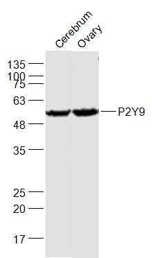
Sample:
Cerebrum (Mouse) Lysate at 40 ug
Ovary (Mouse) Lysate at 40 ug
Primary: Anti-P2Y9 (SL12074R) at 1/1000 dilution
Secondary: IRDye800CW Goat Anti-Rabbit IgG at 1/20000 dilution
Predicted band size: 42 kD
Observed band size: 57 kD
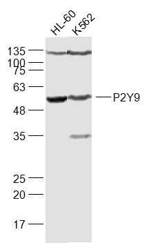
Sample:
HL-60(Human) Cell Lysate at 30 ug
K562(Human) Cell Lysate at 30 ug
Primary: Anti-P2Y9 (SL12074R) at 1/1000 dilution
Secondary: IRDye800CW Goat Anti-Rabbit IgG at 1/20000 dilution
Predicted band size: 42 kD
Observed band size: 57 kD
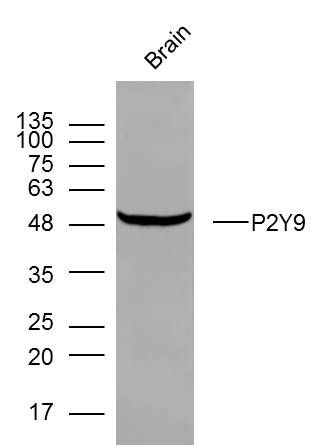
Sample: Brain (mouse) Lysate at 40 ug
Primary: Anti- P2Y9 (SL12074R) at 1/300 dilution
Secondary: IRDye800CW Goat Anti-Rabbit IgG at 1/20000 dilution
Predicted band size: 42 kD
Observed band size: 48 kD
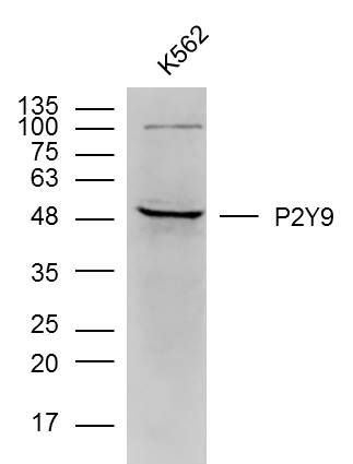
Sample: k562 (human)cell Lysate at 40 ug
Primary: Anti- P2Y9 (SL12074R) at 1/300 dilution
Secondary: IRDye800CW Goat Anti-Rabbit IgG at 1/20000 dilution
Predicted band size: 42 kD
Observed band size: 48 kD
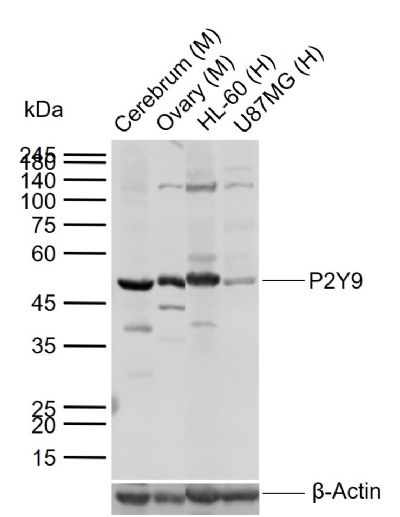
Sample:
Lane 1: Mouse Cerebrum tissue lysates
Lane 2: Mouse Ovary tissue lysates
Lane 3: Human HL60 cell lysates
Lane 4: Human U87MG cell lysates
Primary: Anti-P2Y9 (SL12074R) at 1/1000 dilution
Secondary: IRDye800CW Goat Anti-Rabbit IgG at 1/20000 dilution
Predicted band size: 42 kDa
Observed band size: 50 kDa
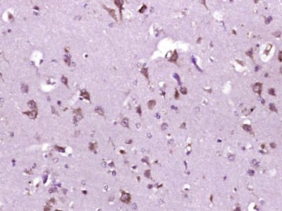
Paraformaldehyde-fixed, paraffin embedded (Human brain glioma); Antigen retrieval by boiling in sodium citrate buffer (pH6.0) for 15min; Block endogenous peroxidase by 3% hydrogen peroxide for 20 minutes; Blocking buffer (normal goat serum) at 37°C for 30min; Antibody incubation with (P2Y9) Polyclonal Antibody, Unconjugated (SL12074R) at 1:400 overnight at 4°C, followed by operating according to SP Kit(Rabbit) (sp-0023) instructionsand DAB staining.
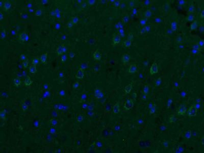
Paraformaldehyde-fixed, paraffin embedded (mouse brain); Antigen retrieval by boiling in sodium citrate buffer (pH6.0) for 15min; Blocking buffer (normal goat serum) at 37°C for 30min; Antibody incubation with (P2Y9) Polyclonal Antibody, Unconjugated (SL12074R) at 1:200 overnight at 4°C, followed by a conjugated Goat Anti-Rabbit IgG antibody (SL0295G-AF488) for 90 minutes, and DAPI for nuclei staining.
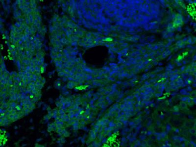
Paraformaldehyde-fixed, paraffin embedded (rat ovary); Antigen retrieval by boiling in sodium citrate buffer (pH6.0) for 15min; Blocking buffer (normal goat serum) at 37°C for 30min; Antibody incubation with (P2Y9) Polyclonal Antibody, Unconjugated (SL12074R) at 1:200 overnight at 4°C, followed by a conjugated Goat Anti-Rabbit IgG antibody (SL0295G-AF488) for 90 minutes, and DAPI for nuclei staining.
|