[IF=7.658] Lei Ma. et al. Identification of the anti-fungal drug fenticonazole nitrate as a novel PPARγ-modulating ligand with good therapeutic index: Structure-based screening and biological validation. Pharmacol Res. 2021 Nov;173:105860 WB ; Mouse.
[IF=5.458] Claire Bryant. et al. Selective Modulator of Nuclear Receptor PPARγ with Reduced Adipogenic Potential Ameliorates Experimental Nephrotic Syndrome. Iscience. 2022 Feb;:104001 WB ; Rat.
[IF=1.88] Shihe Zhang. et al. Astragalus polysaccharide regulates brown adipocytes differentiation by miR-6911 targeting Prdm16. 2021 Nov 05 WB ; Mouse.
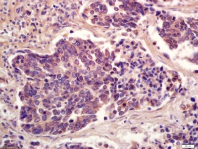
[IF=2.406] Wang, Xu. et al. Modulatory effect of euxanthone in liver cancer-bearing obese mice: crosstalk between PPARγ and TIMP3 signalling axes. 3 Biotech. 2021 Nov;11(11):1-7 IHC ; Mice.
[IF=2.1] Qiankun Quan. et al. Ginsenoside Rg1 reduces β‑amyloid levels by inhibiting CDΚ5‑induced PPARγ phosphorylation in a neuron model of Alzheimer's disease. Mol Med Rep. 2020 Oct;22(4):3277-3288 WB ; Rat.
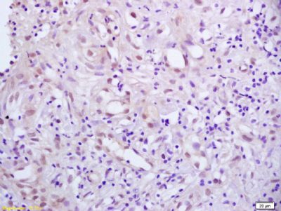
[IF=5.116] Pengyu Hong. et al. Therapeutic potential of small extracellular vesicles derived from lipoma tissue in adipose tissue regeneration—an in vitro and in vivo study. Stem Cell Res Ther. 2021 Dec;12(1):1-13 IHC ; Human.
[IF=4.171] Yingying Tian. et al. Exogenous natural EPA-enriched phosphatidylcholine and phosphatidylethanolamine ameliorate lipid accumulation and insulin resistance via activation of PPARα/γ in mice. Food Funct. 2020 Sep;11(9):8248-8258 WB ; Mouse.
[IF=4.966] Ye Zhanget al. Thymopentin improves the survival of septic mice by promoting the production of 15‐deoxy‐prostaglandin J2 and activating the PPARγ signaling pathway. FASEB J
. 2020 Sep;34(9):11772-11785. WB ; mouse.
[IF=6.133] Huan Y et al. A novel specific PPARγ modulator YR4‐42 ameliorates hyperglycemia and dyslipidemia and hepatic steatosis in diet‐induced obese mice. Diabetes Obes Metab. 2019 Jul 31. WB ; Mouse.
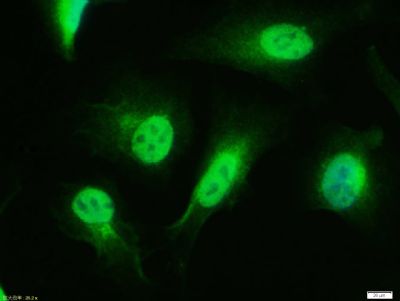
[IF=1.632] Liu Y et al. Isolation and characterization of ovine monocyte-derived macrophages from peripheral blood.(2018)Vet Immunol Immunopathol. Nov;205:83-92. IF ; Sheep.
[IF=3.386] Choung S et al. Treatment with lobeglitazone attenuates hepatic steatosis in diet-induced obese mice.PPAR Res. 2018 Jun 13;2018:4292509. WB ; Mouse.
[IF=0] Yuan, Hai-Feng, et al. "Expression of p-PPARγ in the aging thoracic aorta of spontaneously hypertensive rat and inhibitory effect of rosiglitazone." Asian Pacific Journal of Tropical Biomedicine 4.12 (2014): 977-981. IHSLCP ; Rat.
[IF=4.26] Maganti, Aarthi V., et al. "Peroxisome Proliferator-Activated Receptor-γ Activation Augments the β Cell Unfolded Protein Response and Rescues Early Glycemic Deterioration and β Cell Death in Non-Obese Diabetic Mice."Journal of Biological Chemistry (2016): jbc-M116. WB ; Mouse.
[IF=4.26] Stechschulte, Lance A., et al. "Protein Phosphatase PP5 Controls Bone Mass and the Negative Effects of Rosiglitazone on Bone through Reciprocal Regulation of PPARγ and RUNX2." Journal of Biological Chemistry (2016). WB ; Mouse.
[IF=3.86] Liu, Lei, et al. "Dihydromyricetin delays the onset of hyperglycemia and ameliorates insulin resistance without excessive weight gain in Zucker diabetic fatty rats." Molecular and Cellular Endocrinology (2016). WB ; Rat.
[IF=3.23] Sarr, Ousseynou, et al. "Low birth weight male guinea pig offspring display increased visceral adiposity in early adulthood." PloS one 9.6 (2014): e98433. WB ; Guinea Pig.
[IF=5.58] Agrawal, S., et al. "Pioglitazone Enhances the Beneficial Effects of Glucocorticoids in Experimental Nephrotic Syndrome." Scientific Reports 6 (2016): 24392. IHSLCP ; Rat.
[IF=1.29] Pandurangan, Muthuraman, Jeongeun Park, and Eunjung Kim. "Aspartame downregulates 3T3-L1 differentiation." In Vitro Cellular & Developmental Biology-Animal (2014): 1-7. WB ; Mouse.
[IF=2.233] Kiyoko Maruyama. et al. Indomethacin, a non-steroidal anti-inflammatory drug, induces skin dryness via PPARγ in mice. 2021 Oct 30 WB ; Mouse.
[IF=7.103] Hou et al. CMHX008, a PPARγ partial agonist, enhances insulin sensitivity with minor influences on bone loss. (2018) Genes.Dis. 5:290-299 WB ; Mouse.
[IF=7.422] Dan Wu. et al. A Novel Peroxisome Proliferator-Activated Receptor Gamma Ligand Improves Insulin Sensitivity and Promotes Browning of White Adipose Tissue in Obese Mice. Mol Metab. 2021 Oct;:101363 WB ; Mouse.
[IF=5.059] Stephanie Kimet al. Triphenyl phosphate is a selective PPARγ modulator that does not induce brite adipogenesis in vitro and in vivo. Arch Toxicol
. 2020 Sep;94(9):3087-3103. WB ; mouse.
[IF=1.54] Zhang Y et al. WSF-7 Inhibits Obesity-Mediated PPARγ Phosphorylation and Improves Insulin Sensitivity in 3T3-L1 Adipocytes. Biol Pharm Bull. 2020;43(3):526-532. WB ; mouse.
[IF=7.199] Zhang C et al. Osteoprotegerin Promotes Liver Steatosis by Targeting the ERK-PPARγ-CD36 Pathway. Diabetes. 2019 Jul 10. pii: db181055. WB ; Mouse.
[IF=3.514] Li B et al. Resistin up-regulates LPL expression through the PPARγ-dependent PI3K/AKT signaling pathway impacting lipid accumulation in RAW264. 7 macrophages.Cytokine. 2019 Jul;119:168-174. WB ; Mouse.
[IF=4.5] Liu, Lei, et al. "Dihydromyricetin enhances glucose uptake by inhibition of MEK/ERK pathway and consequent down‐regulation of phosphorylation of PPARγ in 3T3‐L1 cells." Journal of Cellular and Molecular Medicine (2017). WB ; Mouse.
[IF=4.26] Alla, Joshua Abd, et al. "Inhibition of G-protein-coupled Receptor Kinase 2 Prevents the Dysfunctional Cardiac Substrate Metabolism in Fatty Acid Synthase Transgenic Mice." Journal of Biological Chemistry 291.6 (2016): 2583-2600. WB ; Mouse.
[IF=5.08] Liu, Chang, et al. "Identification of a novel selective agonist of PPARγ with no promotion of adipogenesis and less inhibition of osteoblastogenesis." Scientific Reports 5 (2015). WB ; Mouse.
[IF=3.73] Kolli, Vipula, et al. "Partial Agonist, Telmisartan, Maintains PPARγ Serine 112 Phosphorylation, and Does Not Affect Osteoblast Differentiation and Bone Mass." PLOS ONE 9.5 (2014): e96323. WB ; Mouse.