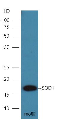
[IF=4.784] Han Y et al. Inonotus obliquus polysaccharides protect against Alzheimer's disease by regulating Nrf2 signaling and exerting antioxidative and antiapoptotic effects.Int J Biol Macromol. 2019 Jun 15;131:769-778. WB ; Mouse.
[IF=6.291] Shaofeng Wu. et al. The neuroprotective effect of curcumin against ATO triggered neurotoxicity through Nrf2 and NF-κB signaling pathway in the brain of ducks. Ecotox Environ Safe. 2021 Dec;228:112965 WB ; Duck.
[IF=6.244] Xinrui Zhang. et al. Wan-Nian-Qing, a Herbal Composite Prescription, Suppresses the Progression of Liver Cancer in Mice by Regulating Immune Response. Front Oncol. 2021; 11: 696282 WB ; Mouse.
[IF=4.872] Shaofeng Wu. et al. Protective effects of curcumin on ATO-induced nephrotoxicity in ducks in relation to suppressed autophagy, apoptosis and dyslipidemia by regulating oxidative stress. Ecotox Environ Safe. 2021 Aug;219:112350 WB ; Duck.
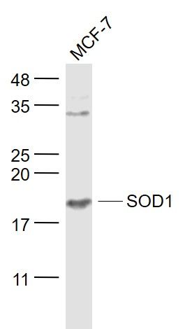
[IF=9.986] Jiyoung Hwang. et al. SOD1 suppresses pro-inflammatory immune responses by protecting against oxidative stress in colitis. Redox Biol. 2020 Oct;37:101760 WB ; Human.
[IF=4.868] Wang Z et al. Aronia melanocarpa Prevents Alcohol-Induced Chronic Liver Injury via Regulation of Nrf2 Signaling in C57BL/6 Mice. Oxid Med Cell Longev. 2020 Jan 8;2020:4054520. WB ; Mouse.
[IF=4.868] Meng B et al. Hepatoprotective Effects of Morchella esculenta against Alcohol-Induced Acute Liver Injury in the C57BL/6 Mouse Related to Nrf-2 and NF-κB Signaling. Oxidative Medicine and Cellular Longevity.2019, Article ID 6029876. WB ; Mouse.
[IF=3.345] Xu et al. Morphological and proteomic analyses reveal that unsaturated guluronate oligosaccharide modulates multiple functional pathways in murine macrophage RAW264.7 cells. (2015) Mar.Drug. 13:1798-818 WB ; Mouse.
[IF=1.69] Han, Ronghui, et al. "Astragalus polysaccharide ameliorates H2O2-induced human umbilical vein endothelial cell injury." Molecular Medicine Reports15.6 (2017): 4027-4034. WB ; Human.
[IF=3.503] Yongfeng Zhang. et al. Aucubin slows the development of osteoporosis by inhibiting osteoclast differentiation via the nuclear factor erythroid 2-related factor 2-mediated antioxidation pathway. Pharm Biol. 2021;59(1):1556-1565 WB ; Mouse.
[IF=3.503] Zhang-Jie Yang. et al. SIRT 3 was involved in Lycium barbarum seed oil protection testis from oxidative stress: in vitro and in vivo analyses. Pharm Biol. 2021;59(1):1314-1325 WB ; Rat.
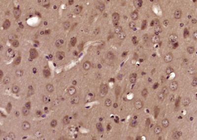
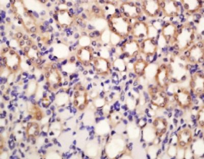
[IF=2.629] Setyaningsih Wiwit A. W.. et al. Ethanolic Extract of Centella asiatica Treatment in the Early Stage of Hyperglycemia Condition Inhibits Glomerular Injury and Vascular Remodeling in Diabetic Rat Model. Evid-Based Compl Alt. 2021;2021:6671130 WB,IHC ; Rat.
[IF=6.551] Yukun Fang. et al. Activation of the ROS/HO-1/NQO1 signaling pathway contributes to the copper-induced oxidative stress and autophagy in duck renal tubular epithelial cells. Sci Total Environ. 2021 Feb;757:143753 WB ; Duck.
[IF=2.323] Liu R et al. Effect of Dietary Methionine Deficiency Followed by a Re-Feeding Phase on the Hepatic Antioxidant Activities of LambsAnimals (Basel).2020 Dec 23;11(1):7. WB ; Lamb.
[IF=2.197] Arfian N et al. Chlorogenic Acid Attenuates Kidney Ischemic/Reperfusion Injury via Reducing Inflammation, Tubular Injury, and Myofibroblast Formation. BioMed Research International, 2019, 1–10. IHSLCP ; Mouse.