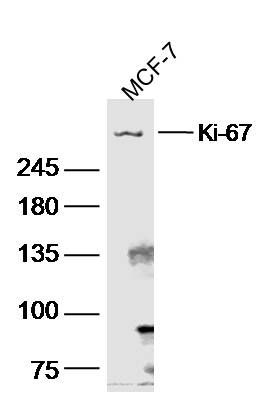
[IF=3.337] Hongzhong Cheng. et al. Circular RNA circLPAR3 Facilitates Esophageal Squamous Cell Carcinoma Progression Through Upregulating HMGB1 via Sponging miR-375/miR-433. Oncotargets Ther. 2020; 13: 7759–7771 WB ; Human.
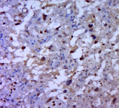
[IF=6.529] Hao Li. et al. Dominant negative TGF-β receptor type II in T lymphocytes promotes anti-tumor immunity by modulating T cell subsets and enhancing CTL responses. Biomed Pharmacother. 2022 Apr;148:112754 IHC ; Mouse.
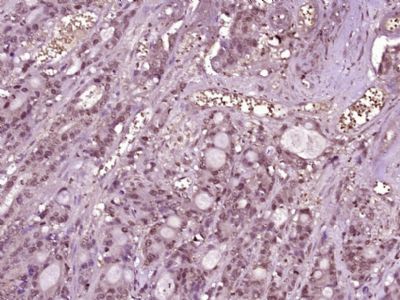
[IF=4.275] Haibin Zhu. et al. Human HAND1 Inhibits the Conversion of Cholesterol to Steroids in Trophoblasts. J Genet Genomics. 2021 Aug;: IHC ; Human.
[IF=2.945] Zhang, Zilong. et al. Circ_FBLN1 promotes the proliferation and osteogenic differentiation of human bone marrow-derived mesenchymal stem cells by regulating let-7i-5p/FZD4 axis and Wnt/β-catenin pathway. 2021 Aug 23 WB ; Human.
[IF=2.248] Wei Shunying. et al. Overexpression of circ_CELSR1 facilitates paclitaxel resistance of ovarian cancer by regulating miR-149-5p/SIK2 axis. Anti-Cancer Drug. 2021 Jun;32(5):496-507 WB ; Human.
[IF=3.565] Liu J. et al. Moxidectin induces Cytostatic Autophagic Cell Death of Glioma Cells through inhibiting the AKT/mTOR Signalling Pathway.. J Cancer. 2020 Aug;11(19):5802-5811 IHC ; Mouse.
[IF=3.647] Liuxian Ban et al. Anti-fungal drug itraconazole exerts anti-cancer effects in oral squamous cell carcinoma via suppressing Hedgehog pathway. Life Sci. 2020 Aug 1;254:117695. IHC ; Human.
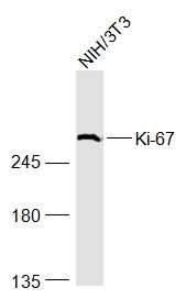
[IF=2.535] Liu J et al. Ivermectin induces autophagy-mediated cell death through the AKT/mTOR signaling pathway in glioma cells. Biosci Rep. 2019 Dec 20;39(12). pii: BSR20192489. WB&IHSLCP ; Human&Mouse.
[IF=3.296] Liang M et al. LncRNA MCM3AP-AS1 promotes proliferation and invasion through regulating miR-211-5p/SPARC axis in papillary thyroid cancer. Endocrine. 2019 Apr 27. IHC ; Human.
[IF=2.705] Ye K et al. Exogenous mesenchymal stem cells affect the function of endogenous lung stem cells (club cells) in phosgene-induced lung injury. Biochem Biophys Res Commun. 2019 Jun 30;514(3):586-592. IHF ; Rat.
[IF=5.595] Yang C et al. MicroRNA-766 promotes cancer progression by targeting NR3C2 in hepatocellular carcinoma. FASEB J. 2019 Jan;33(1):1456-1467. IHSLCP ; Mouse.
[IF=2.645] Xiao H et al. Osthole ameliorates cognitive impairments via augmenting neuronal population in APP/PS1 transgenic mice. Neurosci Res
. 2020 Apr 14;S0168-0102(20)30008-0. ICC,WB ; mouse.
[IF=4.175] Junmin Li. et al. Circ_ZFR contributes to the paclitaxel resistance and progression of non-small cell lung cancer by upregulating KPNA4 through sponging miR-195-5p. Cancer Cell Int. 2021 Dec;21(1):1-15 WB ; Human.
[IF=5.696] Jiang-Tao Fan. et al. Exosomal lncRNA NEAT1 from cancer-associated fibroblasts facilitates endometrial cancer progression via miR-26a/b-5p-mediated STAT3/YKL-40 signaling pathway. Neoplasia. 2021 Jul;23:692 IHC ; Mouse.
[IF=2.754] Wu, Guoxian. et al. Circ-RNF111 aggravates the malignancy of gastric cancer through miR-876-3p-dependent regulation of KLF12. World J Surg Oncol. 2021 Dec;19(1):1-12 IHC ; Human.
[IF=2.363] Wang Li. et al. Trilobatin Induces Apoptosis and Attenuates Stemness Phenotype of Acquired Gefitinib Resistant Lung Cancer Cells via Suppression of NF-κB Pathway. 2021 Apr 16 WB ; Human.
[IF=7.242] Yong Tang. et al. Laminin alpha 4 promotes bone regeneration by facilitating cell adhesion and vascularization. Acta Biomater. 2021 Mar;: IF ; Mouse.
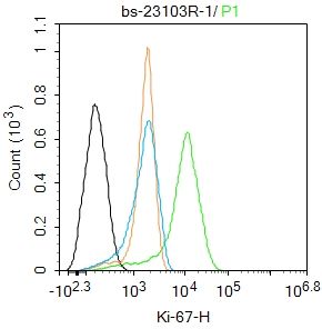
[IF=2.311] Cuina Hanet al. Associations between the expression of SCCA, MTA1, P16, Ki‑67 and the infection of high‑risk HPV in cervical lesions. Oncol Lett
. 2020 Jul;20(1):884-892. IHC ; Human.