The caspase family of cysteine proteases play a key role in apoptosis. Caspase 3 is the most extensively studied apoptotic protein among caspase family members. Caspase 3 is synthesized as inactive pro enzyme that is processed in cells undergoing apoptosis by self proteolysis and/or cleavage by other upstream proteases (e.g. Caspases 8, 9 and 10). The processed form of Caspase 3 consists of large (17kDa) and small (12kDa) subunits which associate to form an active enzyme. Caspase 3 is cleaved at Asp28 Ser29 and Asp175 Ser176. The active Caspase 3 proteolytically cleaves and activates other caspases (e.g. Caspases 6, 7 and 9), as well as relevant targets in the cells (e.g. PARP and DFF). Alternative splicing of this gene results in two transcript variants which encode the same protein. In immunohistochemical studies Caspase 3 expression has been shown to be widespread but not present in all cell types (e.g. commonly reported in epithelial cells of skin, renal proximal tubules and collecting ducts). Differences in the level of Caspase 3 have been reported in cells of short lived nature (eg germinal centre B cells) and those that are long lived (eg mantle zone B cells). Caspase 3 is the predominant caspase involved in the cleavage of amyloid beta 4A precursor protein, which is associated with neuronal death in Alzheimer's disease.
Reacts with Caspase-3 subunit p17 and precursor.
Function:
Involved in the activation cascade of caspases responsible for apoptosis execution. At the onset of apoptosis it proteolytically cleaves poly(ADP-ribose) polymerase (PARP) at a '216-Asp-|-Gly-217' bond. Cleaves and activates sterol regulatory element binding proteins (SREBPs) between the basic helix-loop-helix leucine zipper domain and the membrane attachment domain. Cleaves and activates caspase-6, -7 and -9. Involved in the cleavage of huntingtin. Triggers cell adhesion in sympathetic neurons through RET cleavage.
Subunit:
Heterotetramer that consists of two anti-parallel arranged heterodimers, each one formed by a 17 kDa (p17) and a 12 kDa (p12) subunit. Interacts with BIRC6/bruce.
Subcellular Location:
Cytoplasm.
Tissue Specificity:
Highly expressed in lung, spleen, heart, liver and kidney. Moderate levels in brain and skeletal muscle, and low in testis. Also found in many cell lines, highest expression in cells of the immune system.
Post-translational modifications:
Cleavage by granzyme B, caspase-6, caspase-8 and caspase-10 generates the two active subunits. Additional processing of the propeptides is likely due to the autocatalytic activity of the activated protease. Active heterodimers between the small subunit of caspase-7 protease and the large subunit of caspase-3 also occur and vice versa.
S-nitrosylated on its catalytic site cysteine in unstimulated human cell lines and denitrosylated upon activation of the Fas apoptotic pathway, associated with an increase in intracellular caspase activity. Fas therefore activates caspase-3 not only by inducing the cleavage of the caspase zymogen to its active subunits, but also by stimulating the denitrosylation of its active site thiol.
Similarity:
Belongs to the peptidase C14A family.
SWISS:
P42574
Gene ID:
836
Database links:
Entrez Gene: 836 Human
Entrez Gene: 12367 Mouse
Entrez Gene: 397244 Pig
Entrez Gene: 100008168 Rabbit
Entrez Gene: 25402 Rat
Omim: 600636 Human
SwissProt: P42574 Human
SwissProt: P70677 Mouse
SwissProt: Q95ND5 Pig
SwissProt: Q8MJC3 Rabbit
SwissProt: P55213 Rat
Unigene: 141125 Human
Unigene: 3885 Mouse
Unigene: 10562 Rat
Caspase3广泛分布于各种不同类型的细胞中,是Caspase家族中最重要的凋亡执行者之一,激活的Caspase-3能使许多与细胞结构、细胞周期及DNA修复等相关蛋白或激酶失活,从而使细胞凋亡.
| Picture |
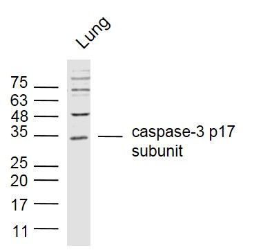
Sample:
Lung (Mouse) Lysate at 40 ug
Primary: Anti- caspase-3 p17 subunit (SL20364R) at 1/300 dilution
Secondary: IRDye800CW Goat Anti-Rabbit IgG at 1/20000 dilution
Predicted band size: 17/32 kD
Observed band size: 17/32 kD
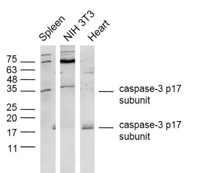
Sample:
Spleen (Mouse) Lysate at 40 ug
NIH/3T3 (Mouse) Cell Lysate at 30 ug
Heart (Mouse) Lysate at 40 ug
Primary: Anti- caspase-3 p17 subunit (SL20364R) at 1/300 dilution
Secondary: IRDye800CW Goat Anti-Rabbit IgG at 1/20000 dilution
Predicted band size: 17/32 kD
Observed band size: 17/32 kD
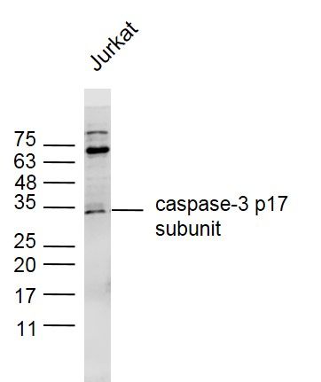
Sample:
Jurkat (human) Cell Lysate at 30 ug
Primary: Anti- caspase-3 p17 subunit (SL20364R) at 1/300 dilution
Secondary: IRDye800CW Goat Anti-Rabbit IgG at 1/20000 dilution
Predicted band size: 17/32 kD
Observed band size: 17/32 kD
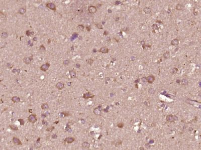
Paraformaldehyde-fixed, paraffin embedded (Human glioma); Antigen retrieval by boiling in sodium citrate buffer (pH6.0) for 15min; Block endogenous peroxidase by 3% hydrogen peroxide for 20 minutes; Blocking buffer (normal goat serum) at 37°C for 30min; Antibody incubation with (caspase-3 p17 subunit) Polyclonal Antibody, Unconjugated (SL20364R) at 1:400 overnight at 4°C, followed by operating according to SP Kit(Rabbit) (sp-0023) instructionsand DAB staining.
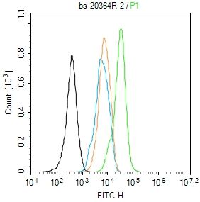
Blank control (blue line): Hela (fixed with 80% methanol (5 min at -20℃) and then permeabilized with 0.1% PBS-Tween for 20 min at room temperature).
Primary Antibody (green line): Rabbit Anti-caspase-3 p12 subunit antibody (SL20364R),Dilution: 1μg /10^6 cells;
Isotype Control Antibody (orange line): Rabbit IgG .
Secondary Antibody (white blue line): Goat anti-rabbit IgG-FITC,Dilution: 1μg /test.
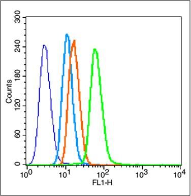
Overlay histogram showing HL 60 cells stained with SL20364R (Green line).
The cells were fixed with 90% methanol (5 min) and then permeabilized with 0.01M PBS-Tween for 20 min. The cells were then incubated in 1x PBS / 10% normal goat serum to block non-specific protein-protein interactions followed by the antibody (SL20364R,1μg/1x10^6 cells) for 30 min at 22℃. The secondary antibody used was fluorescein isothiocyanate goat anti-rabbit IgG (H+L) (SL 0295G-FITC , Brillant blue line) at 1/200 dilution for 30 min at 22℃. Isotype control antibody was rabbit IgG (polyclonal,SL0295P,Orange line) (1μg/1x10^6 cells) used under the same conditions. Unlabelled sample (blue line) was also used as a control. Acquisition of 20,000 events were collected using a 20mW Argon ion laser (488nm) and 525/30 bandpass filter.
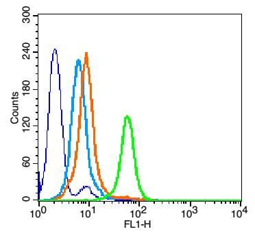
Blank control: NIH/3T3.
Primary Antibody (green line): Rabbit Anti-caspase-3 p17 subunit antibody (SL20364R)
Dilution: 1μg /10^6 cells;
Isotype Control Antibody (orange line): Rabbit IgG .
Secondary Antibody : Goat anti-rabbit IgG-AF488
Dilution: 1μg /test.
Protocol
The cells were fixed with 4% PFA (10min at room temperature)and then permeabilized with 90% ice-cold methanol for 20 min at -20℃. The cells were then incubated in 5%BSA to block non-specific protein-protein interactions for 30 min at room temperature .Cells stained with Primary Antibody for 30 min at room temperature. The secondary antibody used for 40 min at room temperature. Acquisition of 20,000 events was performed.
|
|
|