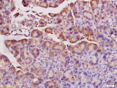
[IF=4.225] Gao Mengyu. et al. The Role of Nrf2 in the PM-Induced Vascular Injury Under Real Ambient Particulate Matter Exposure in C57/B6 Mice. Front Pharmacol. 2021 Feb;12:206 IHC ; Mouse.
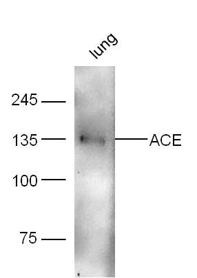
[IF=1.789] Xiaohua Liang . et al. Vitamin A deficiency indicating as low expression of LRAT may be a novel biomarker of primary hypertension. Clin Exp Hypertens. 2021;43(2):151-163 WB ; Human.
[IF=3.448] Chen ZZ et al. Tanshinone IIA contributes to the pathogenesis of endometriosis via renin angiotensin system by regulating the dorsal root ganglion axon sprouting. Life Sci. 2019 Nov 20;48:117085. IHSLCP&WB ; Rat.
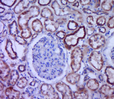
[IF=3.166] Deng T et al. Exposure to diisononyl phthalate induced an increase in blood pressure through activation of the ACE/ AT1R axis and inhibition of NO production. Toxicol Lett. 2019 Jul;309:42-50. IHC ; Mouse.
[IF=3.373] Zhang et al. Simultaneous targeting of ATM and Mcl-1 increases cisplatin sensitivity of cisplatin-resistant non-small cell lung cancer. (2017) Cancer.Biol.Ther. 18:606-615 WB ; Human.
[IF=3.263] Ademola Adetokunbo Oyagbemi et al. Antihypertensive power of Naringenin is mediated via attenuation of mineralocorticoid receptor (MCR)/ angiotensin converting enzyme (ACE)/ kidney injury molecule (Kim-1) signaling pathway. Eur J Pharmacol. 2020 Aug 5;176:173142. IHC ; Rat.
[IF=1.984] Liu H et al. Ginsenoside Rg3 Attenuates Angiotensin II-Mediated Renal Injury in Rats and Mice by Upregulating Angiotensin-Converting Enzyme 2 in the Renal Tissue. Evid Based Complement Alternat Med. 2019 Nov 29;2019:6741057. IHSLCP ; Rat.
[IF=4.358] Deng T et al. Di-(2-ethylhexyl) phthalate induced an increase in blood pressure via activation of ACE and inhibition of the bradykinin-NO pathway. Environ Pollut. 2019 Apr;247:927-934. IHC ; Mouse.
[IF=3.974] Xie X et al. Comparing the effects of diethylhexyl phthalate and dibutyl phthalate exposure on hypertension in mice.Ecotoxicol Environ Saf. 2019 Jun 15;174:75-82. IHC ; Mouse.
[IF=1.26] Jiang et al. Ginsenoside Rg3 induces ginsenoside Rb1-comparable cardioprotective effects independent of reducing blood pressure in spontaneously hypertensive rats. (2017) Exp.Ther.Med. 14:4977-4985 IHSLCP ; Rat.
[IF=1.92] Liu, Chen, et al. "Pulmonary artery denervation improves pulmonary arterial hypertension induced right ventricular dysfunction by modulating the local renin-angiotensin-aldosterone system." BMC Cardiovascular Disorders 16.1 (2016): 192. WB ; Dog.