[IF=1.69] Zhao, Zhan‐Zheng, et al. "Effects of recombinant human endostatin on peritoneal angiogenesis in peritoneal dialysis rats." Nephrology 16.6 (2011): 599-606. Rat.
[IF=7.991] Xiangfeng Li. et al. Design of macropore structure and micro-nano topography to promote the early neovascularization and osteoinductivity of biphasic calcium phosphate bioceramics. Mater Design. 2022 Apr;216:110581 IF ; Human.
[IF=4.379] Zhou, Liuhua. et al. Effect of uncultured adipose-derived stromal vascular fraction on preventing urethral stricture formation in rats. Sci Rep-Uk. 2022 Mar;12(1):1-10 WB ; Rat.
[IF=5.396] Ali Bagherian. et al. Anti-glioblastoma effects of nanomicelle-curcumin plus erlotinib. Food Funct. 2021 Oct;: WB ; Human.
[IF=6.832] Liu, Jingyu. et al. Therapeutic effect of adipose stromal vascular fraction spheroids for partial bladder outlet obstruction induced underactive bladder. Stem Cell Res Ther. 2022 Dec;13(1):1-16 WB ; Rat.
[IF=5.396] Wenji Hu. et al. Structural characterization of polysaccharide purified from Amanita caesarea and its pharmacological basis for application in Alzheimer's disease: endoplasmic reticulum stress. Food Funct. 2021 Oct;: WB ; Human.
[IF=1.94] De Nan. et al. In Vitro Study of Adipose-Derived Mesenchymal Stem Cells Transduced with Lentiviral Vector Carrying the Brain-Derived Neurotrophic Factor Gene. Int J Stem Cells. 2020; 13(3): 386–393 WB ; Mouse.
[IF=5.097] Yan L et al. Exosomes produced from 3D cultures of umbilical cord mesenchymal stem cells in a hollow-fiber bioreactor show improved osteochondral regeneration activity. Cell Biology and Toxicology. WB ; Human.
[IF=-] YANG JW. Chondrocyte extracellular matrix-derived peptide. US 2019 / 0111112 A1 IHSLCP ; rabbit.
[IF=4.929] Zhou L et al.Protective Effects of Uncultured Adipose‐Derived Stromal Vascular Fraction on Testicular Injury Induced by Torsion‐Detorsion in Rats. (2018) Stem Cells Translational Medicine WB ; Rat.
[IF=2.959] Peng W et al.Expression of osteoprotegerin and receptor activator for the nuclear factor-κB ligand in XACB/LSLVbFGF/MSCs transplantation for repair of rabbit femoral head defect necrosis.(2018) J. Cell. Biochem. Oct 18 WB ; Rabbit.
[IF=2.28] Liu et al. B-cell leukemia/lymphoma 10 promotes angiogenesis in an experimental corneal neovascularization model. (2018) Eye.(Lond). FC/FACS ; Mouse.
[IF=2.478] Liu G et al. B-cell leukemia/lymphoma 10 promotes angiogenesis in an experimental corneal neovascularization model. Eye,2018 32(7), 1220–1231. FCM ; Mouse.
[IF=5.62] He, Ting, et al. "Tumor cell-secreted angiogenin induces angiogenic activity of endothelial cells by suppressing miR-542-3p." Cancer Letters (2015). WB ; Human.
[IF=1.96] Turgut, Burak, et al. "Impact of trastuzumab on wound healing in experimental glaucoma surgery." Clinical & Experimental Ophthalmology (2014). IHSLCP ; Rabbit.
[IF=1.93] Lee, Hye Sook, et al. "Anti-neovascular effect of chondrocyte-derived extracellular matrix on corneal alkaline burns in rabbits." Graefes Archive for Clinical and Experimental Ophthalmology (2014): 1-11. IHSLCP ; Rabbit.
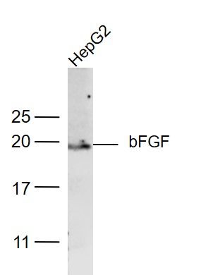
[IF=1.69] Gao, Dan, et al. "Effect of peritoneal dialysis on expression of vascular endothelial growth factor, basic fibroblast growth factor and endostatin of the peritoneum in peritoneal dialysis patients." Nephrology 16.8 (2011): 736-742. WB ; Human.
[IF=1.64] Eren, Kenan, et al. "The Suppression of Wound Healing Response with Sirolimus and Sunitinib Following Experimental Trabeculectomy in a Rabbit Model." Current Eye Research (2016): 1-10. IHSLCP ; Rabbit.
[IF=3.514] Liuhong Liu. et al. Experimental study on the effect of chrysin on skin injury induced by amiodarone extravasation in rats. Microvasc Res. 2022 Jan;139:104257 IHC ; rat.
[IF=1.994] YING-HAO HAN. et al. Peroxiredoxin II Inhibits Alcohol-induced Apoptosis in L02 Hepatocytes Through AKT/β-Catenin Signaling Pathway. Anticancer Res. 2020 Aug;40(8):4491-4504 WB ; Human.
[IF=4.65] Pan, Ruo-Lang, et al. "Delta-like 1 serves as a new target and contributor to liver fibrosis down-regulated by mesenchymal stem cell transplantation." Journal of Biological Chemistry 286.14 (2011): 12340-12348. Mouse.
[IF=3.571] Mei F et al. Collagen peptides isolated from Salmon salar and Tilapia nilotica skin accelerate wound healing by altering cutaneous microbiome colonization via up-regulated NOD2 and BD14. Agric. Food Chem. 2020, 68, 6, 1621-1633. IHSLCP ; Rat.
[IF=2.247] Tort S et al. The effect of a new wound dressing on wound healing: Biochemical and histopathological evaluation. Burns. 2019 Dec 17. pii: S0305-4179(18)30976-8. IHSLCP ; Rat.
[IF=0.181] H Mu et al. HCMSLVencoded IE2 induces anxiety-depression and cognitive impairment in UL122 genetically-modified mice. Int J Clin Exp Pathol 2019;12(11):4087-4095. WB&IHSLCP ; Mouse.
[IF=2.136] Gao XX et al.
Effects of L-arginine on endometrial microvessel density in nutrient-restricted Hu sheep.Theriogenology. 2018 Oct 1;119:252-258. IHSLCP&WB ; Sheep.
[IF=0.37] Shkurupy, V. A., et al. "In Vitro Effects of Nanosized Diamond Particles on Macrophages." Bulletin of Experimental Biology and Medicine (2015): 1-4. Mouse.
[IF=2.83] Sun, Fei, et al. "Structural integrity, immunogenicity and biomechanical evaluation of rabbit decelluarized tracheal matrix." Journal of Biomedical Materials Research Part A (2014). IHSLCP ; Rabbit.
[IF=1.93] Chen, Bo-Yu, et al. "Altered TGF-β2 and bFGF expression in scleral desmocytes from an experimentally-induced myopia guinea pig model." Graefes Archive for Clinical and Experimental Ophthalmology (2013): 1-12. Pig, Guinea Pig.
[IF=2.89] Liu, Kai-Jun, et al. "Analysis of olfactory ensheathing glia transplantation-induced repair of spinal cord injury by electrophysiological, behavioral, and histochemical methods in rats." Journal of molecular neuroscience 41.1 (2010): 25-29. Human.
[IF=3.49] Turgut, Burak, et al. "Topical infliximab for the suppression of wound healing following experimental glaucoma filtration surgery." Drug Design, Development and Therapy 8 (2014): 421-429. IHSLCP ; Rabbit.