[IF=-] Kwon,et al.Luterion and separating and culturing methods for same.(2017) . :. IF ; Plant.
[IF=1.55] Jia, Lianqun., et al. "Effects of Tanshinone IIA on the modulation of miR‑33a and the SREBP‑2/Pcsk9 signaling pathway in hyperlipidemic rats." Molecular Medicine Reports (2016). IHSLCP ; Rat.
[IF=5.34] Wei Zou. et al. Imperatae rhizoma-Hedyotis diffusa Willd. herbal pair alleviates nephrotic syndrome by integrating anti-inflammatory and hypolipidaemic effects. Phytomedicine. 2021 Sep;90:153644 WB ; Rat.
[IF=2.74] Kyoung Jin Leeet al. Real-time monitoring of oncolytic VSV properties in a novel in vitro microphysiological system containing 3D multicellular tumor spheroids. PLoS One
. 2020 Jul 6;15(7):e0235356. IF ; Human.
[IF=2.629] Mei Hui. et al. The Hypolipidemic Effect of Dalbergia odorifera T. C. Chen Leaf Extract on Hyperlipidemic Rats and Its Mechanism Investigation Based on Network Pharmacology. Evid-Based Compl Alt. 2021;2021:3155266 WB ; Rat.
[IF=4.268] Grube,et al.Mining the Secretome of C2C12 Muscle Cells: Data Dependent Experimental Approach To Analyze Protein Secretion Using Label-Free Quantification and Peptide Based Analysis.(2018) Journal of Proteome Research. 17:879-890. WB ; Mouse.
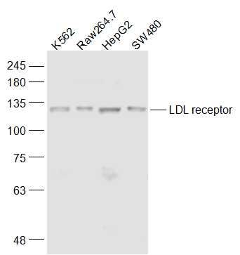
[IF=2.7] Kou, Shuming, et al. "Synergetic cholesterol-lowering effects of main alkaloids from Rhizoma Coptidis in HepG2 cells and hypercholesterolemia hamsters." Life Sciences (2016). WB ; Human.
[IF=2.97] Wu, Hao, et al. "The antihypercholesterolemic effect of jatrorrhizine isolated from Rhizoma Coptidis." Phytomedicine (2014). WB ;