This intronless gene encodes a 70kDa heat shock protein which is a member of the heat shock protein 70 family. In conjuction with other heat shock proteins, this protein stabilizes existing proteins against aggregation and mediates the folding of newly translated proteins in the cytosol and in organelles. It is also involved in the ubiquitin-proteasome pathway through interaction with the AU-rich element RNA-binding protein 1. The gene is located in the major histocompatibility complex class III region, in a cluster with two closely related genes which encode similar proteins. [provided by RefSeq, Jul 2008].
Function:
In cooperation with other chaperones, Hsp70s stabilize preexistent proteins against aggregation and mediate the folding of newly translated polypeptides in the cytosol as well as within organelles. These chaperones participate in all these processes through their ability to recognize nonnative conformations of other proteins. They bind extended peptide segments with a net hydrophobic character exposed by polypeptides during translation and membrane translocation, or following stress-induced damage. In case of rotavirus A infection, serves as a post-attachment receptor for the virus to facilitate entry into the cell.
Subunit:
Component of the CatSper complex. Identified in a mRNP granule complex, at least composed of ACTB, ACTN4, DHX9, ERG, HNRNPA1, HNRNPA2B1, HNRNPAB, HNRNPD, HNRNPL, HNRNPR, HNRNPU, HSPA1, HSPA8, IGF2BP1, ILF2, ILF3, NCBP1, NCL, PABPC1, PABPC4, PABPN1, RPLP0, RPS3, RPS3A, RPS4X, RPS8, RPS9, SYNCRIP, TROVE2, YBX1 and untranslated mRNAs. Interacts with TSC2. Interacts with IRAK1BP1. Interacts with TERT; the interaction occurs in the absence of the RNA component, TERC, and dissociates once the TERT complex has formed. Interacts with DNAJC7. Interacts with CHCHD3.
Subcellular Location:
Cytoplasm. Note=Localized in cytoplasmic mRNP granules containing untranslated mRNAs.
Tissue Specificity:
HSPA1B is testis-specific.
Similarity:
Belongs to the heat shock protein 70 family.
SWISS:
P0DMV8
Gene ID:
3303
Database links:
Entrez Gene: 281825 Cow
Entrez Gene: 3303 Human
Entrez Gene: 3304 Human
Entrez Gene: 15511 Mouse
Entrez Gene: 193740 Mouse
Entrez Gene: 24472 Rat
Entrez Gene: 294254 Rat
Omim: 140550 Human
Omim: 603012 Human
SwissProt: Q27975 Cow
SwissProt: P0DMV8 Human
SwissProt: P0DMV9 Human
SwissProt: P17879 Mouse
SwissProt: Q61696 Mouse
SwissProt: Q07439 Rat
Unigene: 27882 Human
Unigene: 719966 Human
Unigene: 728810 Human
Unigene: 1950 Rat
Unigene: 228225 Rat
信号传导(Signaling Intermediates)
HSP-70是细胞受应激原刺激后诱导产生的一组应激蛋白,与肿瘤发生、增殖及分化有关。
环境和病生理性应激原均可导致肌体合成一组应急蛋白既热休克蛋白。许多热休克蛋白,包括HSP70家族成员,均参与蛋白的变性-复性、折叠-解折叠、运输-易位、活化-非活化和分泌等过程。
HSP70与类固醇受体、肌动蛋白、P53等蛋白密切相关。HSP70还参与热应激原、细胞毒药物和其他损伤所引起的应急反应,对机体起一定的保护作用。
| Picture |
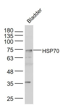
Sample:
Bladder (Mouse) Lysate at 40 ug
Primary: Anti-HSP70 (SL0126R) at 1/300 dilution
Secondary: IRDye800CW Goat Anti-Rabbit IgG at 1/20000 dilution
Predicted band size: 70 kD
Observed band size: 70 kD
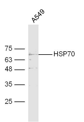
Sample:
A549 Cell (Human) Lysate at 30 ug
Primary: Anti-HSP70 (SL 0126R) at 1/300 dilution
Secondary: IRDye800CW Goat Anti-Rabbit IgG at 1/20000 dilution
Predicted band size: 70 kD
Observed band size: 70 kD
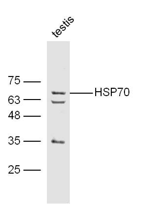
Sample:
Testis (Mouse) Lysate at 40 ug
Primary: Anti-HSP70 (SL 0126R) at 1/300 dilution
Secondary: IRDye800CW Goat Anti-Rabbit IgG at 1/20000 dilution
Predicted band size: 70 kD
Observed band size: 70 kD
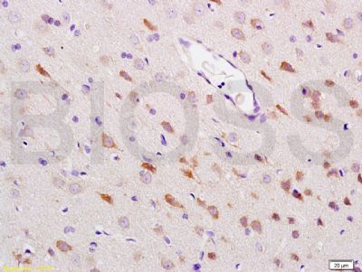
Tissue/cell: rat brain tissue; 4% Paraformaldehyde-fixed and paraffin-embedded;
Antigen retrieval: citrate buffer ( 0.01M, pH 6.0 ), Boiling bathing for 15min; Block endogenous peroxidase by 3% Hydrogen peroxide for 30min; Blocking buffer (normal goat serum,SLC0005) at 37℃ for 20 min;
Incubation: Anti-HSP-70 Polyclonal Antibody, Unconjugated(SL0126R) 1:200, overnight at 4°C, followed by conjugation to the secondary antibody(SP-0023) and DAB(SLC0010) staining
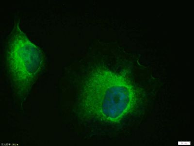
Hela cell; 4% Paraformaldehyde-fixed; Triton X-100 at room temperature for 20 min; Blocking buffer (normal goat serum, SLC0005) at 37°C for 20 min; Antibody incubation with (HSP70) polyclonal Antibody, Unconjugated (SL0126R) 1:100, 90 minutes at 37°C; followed by a conjugated Goat Anti-Rabbit IgG antibody at 37°C for 90 minutes, DAPI (blue, C02-04002) was used to stain the cell nuclei.
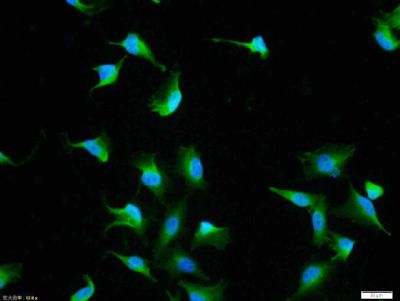
Tissue/cell: A549 cell; 4% Paraformaldehyde-fixed; Triton X-100 at room temperature for 20 min; Blocking buffer (normal goat serum, SLC0005) at 37°C for 20 min; Antibody incubation with (HSP70) polyclonal Antibody, Unconjugated (SL0126R) 1:100, 90 minutes at 37°C; followed by a FITC conjugated Goat Anti-Rabbit IgG antibody at 37°C for 90 minutes, DAPI (blue, C02-04002) was used to stain the cell nuclei.
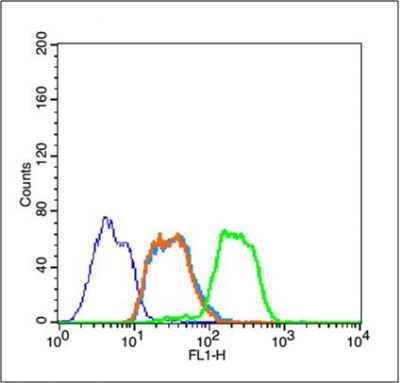
Blank control (blue line): Jurkat (blue).
Primary Antibody (green line): Rabbit Anti-HSP70 antibody (SL0126R)
Dilution: 1μg /10^6 cells;
Isotype Control Antibody (orange line): Rabbit IgG .
Secondary Antibody (white blue line): Goat anti-rabbit IgG-FITC
Dilution: 1μg /test.
Protocol
The cells were fixed with 2% paraformaldehyde (10 min) , then permeabilized with 90% ice-cold methanol for 30 min on ice. Cells stained with Primary Antibody for 30 min at room temperature. The cells were then incubated in 1 X PBS/2%BSA/10% goat serum to block non-specific protein-protein interactions followed by the antibody for 15 min at room temperature. The secondary antibody used for 40 min at room temperature. Acquisition of 20,000 events was performed.
|
|
|