This protein is a member of the paired box (PAX) family of transcription factors. Members of the PAX family typically contain a paired box domain and a paired-type homeodomain. These proteins play critical roles during fetal development. Mutations in paired box gene 3 are associated with Waardenburg syndrome, craniofacial-deafness-hand syndrome, and alveolar rhabdomyosarcoma. The translocation t(2;13)(q35;q14), which represents a fusion between PAX3 and the forkhead gene, is a frequent finding in alveolar rhabdomyosarcoma. Alternative splicing results in transcripts encoding isoforms with different SLCtermini.
Function:
Transcription factor that may regulate cell proliferation, migration and apoptosis. Involved in neural development and myogenesis.
Subunit:
Can bind to DNA as a homodimer or a heterodimer with PAX7. Interacts with PAXBP1; the interaction links PAX3 to a WDR5-containing histone methyltransferase complex. Interacts with DAXX.
Subcellular Location:
Nucleus.
DISEASE:
Waardenburg syndrome 1 (WS1) [MIM:193500]: WS1 is an autosomal dominant disorder characterized by non-progressive sensorineural deafness, pigmentary disturbances such as frontal white blaze of hair, heterochromia of irides, white eyelashes, leukoderma, and wide bridge of nose owing to lateral displacement of the inner canthus of each eye (dystopia canthorum). WS1 shows variable clinical expression and some affected individuals do not manifest hearing impairment or iris pigmentation disturbances. Dystopia canthorum is the most consistent sign and is found in 98% of the patients. Note=The disease is caused by mutations affecting the gene represented in this entry.
Waardenburg syndrome 3 (WS3) [MIM:148820]: WS3 is an autosomal dominant disorder characterized by sensorineural deafness, pigmentary disturbances, dystopia canthorum and limb anomalies such as hypoplasia of the musculoskeletal system, flexion contractures, fusion of the carpal bones, syndactylies. Note=The disease is caused by mutations affecting the gene represented in this entry.
Craniofacial-deafness-hand syndrome (CDHS) [MIM:122176]: Thought to be an autosomal dominant disease which comprises absence or hypoplasia of the nasal bones, hypoplastic maxilla, small and short nose with thin nares, limited movement of the wrist, short palpebral fissures, ulnar deviation of the fingers, hypertelorism and profound sensory-neural deafness. Note=The disease is caused by mutations affecting the gene represented in this entry.
Rhabdomyosarcoma 2 (RMS2) [MIM:268220]: A form of rhabdomyosarcoma, a highly malignant tumor of striated muscle derived from primitive mesenchymal cells and exhibiting differentiation along rhabdomyoblastic lines. Rhabdomyosarcoma is one of the most frequently occurring soft tissue sarcomas and the most common in children. It occurs in four forms: alveolar, pleomorphic, embryonal and botryoidal rhabdomyosarcomas. Note=The gene represented in this entry is involved in disease pathogenesis. A chromosomal aberration involving PAX3 is found in rhabdomyosarcoma. Translocation (2;13)(q35;q14) with FOXO1. The resulting protein is a transcriptional activator.
Note=A chromosomal aberration involving PAX3 is a cause of rhabdomyosarcoma. Translocation t(2;2)(q35;p23) with NCOA1 generates the NCOA1-PAX3 oncogene consisting of the N-terminus part of PAX3 and the SLCterminus part of NCOA1. The fusion protein acts as a transcriptional activator. Rhabdomyosarcoma is the most common soft tissue carcinoma in childhood, representing 5-8% of all malignancies in children.
Similarity:
Belongs to the paired homeobox family.
Contains 1 homeobox DNA-binding domain.
Contains 1 paired domain.
SWISS:
P24610
Gene ID:
5077
Database links:
Entrez Gene: 5077 Human
Entrez Gene: 18505 Mouse
Entrez Gene: 114502 Rat
Omim: 606597 Human
SwissProt: P23760 Human
SwissProt: P24610 Mouse
SwissProt: Q0IH87 Xenopus laevis
SwissProt: Q645N4 Xenopus laevis
Unigene: 42146 Human
Unigene: 1371 Mouse
Unigene: 225252 Rat
同源结构域蛋白(Homeodomain Proteins)
PAX-3属于转录抑制因子。在胚胎发育和肿瘤生长中起关键作用。其突变和某些肿瘤的发病有关。
| Picture |
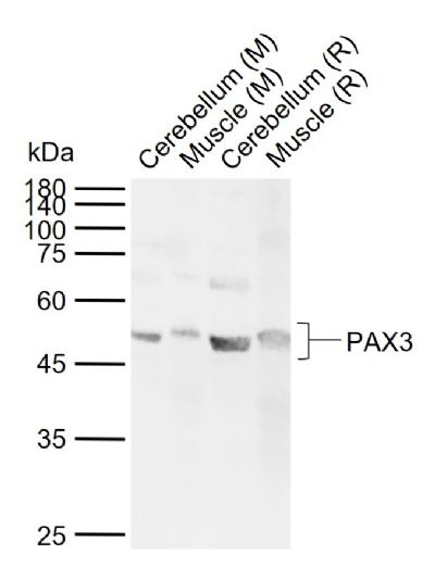
Sample:
Lane 1: Mouse Cerebellum tissue lysates
Lane 2: Mouse Muscle tissue lysates
Lane 3: Rat Cerebellum tissue lysates
Lane 4: Rat Muscle tissue lysates
Primary: Anti-PAX3 (SL1097R) at 1/1000 dilution
Secondary: IRDye800CW Goat Anti-Rabbit IgG at 1/20000 dilution
Predicted band size: 53 kDa
Observed band size: 53 kDa
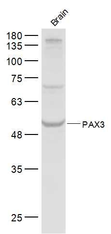
Sample:
Brain (Mouse) Lysate at 40 ug
Primary: Anti-PAX3 (SL1097R) at 1/300 dilution
Secondary: IRDye800CW Goat Anti-Rabbit IgG at 1/20000 dilution
Predicted band size: 53 kD
Observed band size: 53 kD
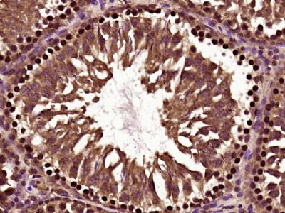
Paraformaldehyde-fixed, paraffin embedded (Rat testis); Antigen retrieval by boiling in sodium citrate buffer (pH6.0) for 15min; Block endogenous peroxidase by 3% hydrogen peroxide for 20 minutes; Blocking buffer (normal goat serum) at 37°C for 30min; Antibody incubation with (PAX3) Polyclonal Antibody, Unconjugated (SL1097R) at 1:400 overnight at 4°C, followed by a conjugated secondary antibody (sp-0023) for 20 minutes and DAB staining.
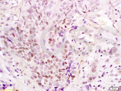
Tissue/cell: Human esophageal carcinoma; 4% Paraformaldehyde-fixed and paraffin-embedded;
Antigen retrieval: citrate buffer ( 0.01M, pH 6.0 ), Boiling bathing for 15min; Block endogenous peroxidase by 3% Hydrogen peroxide for 30min; Blocking buffer (normal goat serum,SLC0005) at 37℃ for 20 min;
Incubation: Anti-PAX3 Polyclonal Antibody, Unconjugated(SL1097R) 1:200, overnight at 4°C, followed by conjugation to the secondary antibody(SP-0023) and DAB(SLC0010) staining
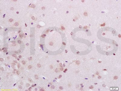
Tissue/cell: rat brain tissue; 4% Paraformaldehyde-fixed and paraffin-embedded;
Antigen retrieval: citrate buffer ( 0.01M, pH 6.0 ), Boiling bathing for 15min; Block endogenous peroxidase by 3% Hydrogen peroxide for 30min; Blocking buffer (normal goat serum,SLC0005) at 37℃ for 20 min;
Incubation: Anti-PAX3 Polyclonal Antibody, Unconjugated(SL1097R) 1:200, overnight at 4°C, followed by conjugation to the secondary antibody(SP-0023) and DAB(SLC0010) staining
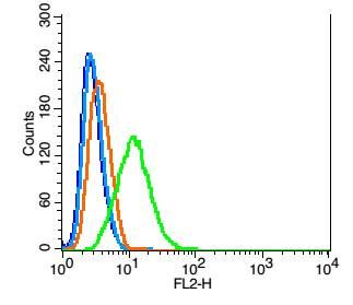
Blank control: Hela(blue).
Primary Antibody:Rabbit Anti- PAX3 antibody(SL1097R), Dilution: 1μg in 100 μL 1X PBS containing 0.5% BSA;
Isotype Control Antibody: Rabbit IgG(orange) ,used under the same conditions );
Secondary Antibody: Goat anti-rabbit IgG-PE(white blue), Dilution: 1:200 in 1 X PBS containing 0.5% BSA.
Protocol
The cells were fixed with 2% paraformaldehyde (10 min) , then permeabilized with 90% ice-cold methanol for 30 min on ice. Antibody (SL1097R, 1μg /1x10^6 cells) were incubated for 30 min on the ice, followed by 1 X PBS containing 0.5% BSA + 1 0% goat serum (15 min) to block non-specific protein-protein interactions. Then the Goat Anti-rabbit IgG/PE antibody was added into the blocking buffer mentioned above to react with the primary antibody of SL1097R at 1/200 dilution for 30 min on ice. Acquisition of 20,000 events was performed.
|
|
|