Microtubules are constituent parts of the mitotic apparatus, cilia, flagella, and elements of the cytoskeleton. They consist principally of 2 soluble proteins, alpha- and beta-tubulin, each of about 55,000 kDa.
Antibodies against beta Tubulin are useful as loading controls for Western Blotting. However it should be noted that levels of beta Tubulin may not be stable in certain cells. For example, expression of tubulin in adipose tissue is very low (Spiegelman and Farmer, Cell, 1982, 29(1):53-60) and therefore beta Tubulin should not be used as loading control for these tissues.
Function:
Tubulin is the major constituent of microtubules. It binds two moles of GTP, one at an exchangeable site on the beta chain and one at a non-exchangeable site on the alpha chain.
Subunit:
Dimer of alpha and beta chains. May interact with RNABP10. Interacts with PIFO. Interacts with MX1.
Subcellular Location:
Cytoplasm, cytoskeleton.
Tissue Specificity:
Ubiquitously expressed with highest levels in spleen, thymus and immature brain.
Post-translational modifications:
Some glutamate residues at the SLCterminus are polyglutamylated. This modification occurs exclusively on glutamate residues and results in polyglutamate chains on the gamma-carboxyl group. Also monoglycylated but not polyglycylated due to the absence of functional TTLL10 in human. Monoglycylation is mainly limited to tubulin incorporated into axonemes (cilia and flagella) whereas glutamylation is prevalent in neuronal cells, centrioles, axonemes, and the mitotic spindle. Both modifications can coexist on the same protein on adjacent residues, and lowering glycylation levels increases polyglutamylation, and reciprocally. The precise function of such modifications is still unclear but they regulate the assembly and dynamics of axonemal microtubules (Probable).
Similarity:
Belongs to the tubulin family.
SWISS:
Q13509
Gene ID:
203068
Database links:
Entrez Gene: 396254 Chicken
Entrez Gene: 203068 Human
Entrez Gene: 380418 Xenopus laevis
Omim: 191130 Human
SwissProt: P07437 Human
SwissProt: P99024 Mouse
SwissProt: Q767L7 Pig
SwissProt: P69897 Rat
SwissProt: Q91575 Xenopus laevis
Unigene: 63696 Human
Unigene: 706187 Human
Unigene: 714425 Human
Unigene: 46552 Xenopus laevis
结构蛋白(Structural Proteins)
tubulin是一种大量存在于哺乳动物脑组织中的微管亚基蛋白,在结构上是由两个极为相近的α和β亚基组成的二聚体、多聚体形成微管细丝,是微管的主要成分。
微管蛋白是球形分子, 有两种类型:α微管蛋白(α-tubulin)货号:SL0195R和β微管蛋白(β-tubulin), 这两种微管蛋白具有相似的三维结构, 能够紧密地结合成二聚体, 作为微管组装的亚基。 α亚基由450个氨基酸组成, β亚基是由455个氨基酸组成, 这两种亚基有35~40%的氨基酸序列同源, 表明编码它们的基因可能是由同一原始祖先演变而来.
| Picture |
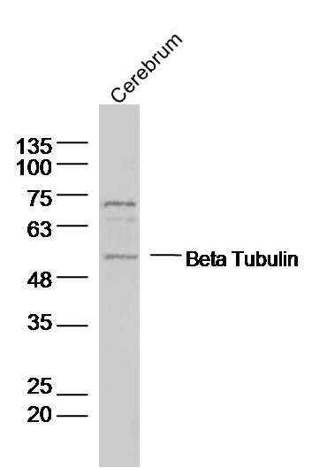
Sample:
Cerebrum (Rat) Lysate at 40 ug
Primary: Anti- Beta Tubulin (SL0715R) at 1/300 dilution
Secondary: IRDye800CW Goat Anti-Rabbit IgG at 1/20000 dilution
Predicted band size: 50 kD
Observed band size: 50 kD
Sample: Cerebrum (Mouse) Lysate at 40 ug
Primary: Anti-Beta Tubulin (SL0715R) at 1/300 dilution
Secondary: IRDye800CW Goat Anti-Rabbit IgG at 1/20000 dilution
Predicted band size: 50 kD
Observed band size: 50 kD
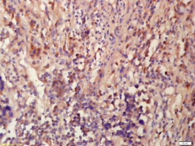
Tissue/cell: human lung carcinoma; 4% Paraformaldehyde-fixed and paraffin-embedded;
Antigen retrieval: citrate buffer ( 0.01M, pH 6.0 ), Boiling bathing for 15min; Block endogenous peroxidase by 3% Hydrogen peroxide for 30min; Blocking buffer (normal goat serum,SLC0005) at 37∩ for 20 min;
Incubation: Anti-Tubulin-Beta Polyclonal Antibody, Unconjugated(SL0715R) 1:200, overnight at 4∑C, followed by conjugation to the secondary antibody(SP-0023) and DAB(SLC0010) staining
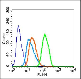
Blank control (blue line): Hela
Primary Antibody (green line): Rabbit Anti-Beta Tubulin antibody (SL0715R)
Dilution: 1μg /10^6 cells;
Isotype Control Antibody (orange line): Rabbit IgG .
Secondary Antibody (white blue line): Goat anti-rabbit IgG-FITC
Dilution: 1μg /test.
Protocol
The cells were fixed with 70% ethanol (Overnight at 4℃) and then permeabilized with 90% ice-cold methanol for 30 min on ice. Cells stained with Primary Antibody for 30 min at room temperature. The cells were then incubated in 1 X PBS/2%BSA/10% goat serum to block non-specific protein-protein interactions followed by the antibody for 15 min at room temperature. The secondary antibody used for 40 min at room temperature. Acquisition of 20,000 events was performed.
|
|
|