Specific References (6) | SL1309R has been referenced in 6 publications.
[IF=1.396] Xiong Y et al. Functions of T-cell subsets and their related cytokines in the pathological processes of autoimmune encephalomyelitic mice.(2018) Int J Clin Exp Pathol;11(10):4817-4826. FC&IHC ; Mouse.
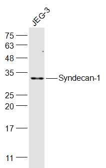
[IF=5.733] Jiang Yufei. et al. Osteoprotegerin interacts with syndecan-1 to promote human endometrial stromal decidualization by decreasing Akt phosphorylation. Hum Reprod. 2020 Nov;35(11):2439-2453 IP ; Human.
[IF=4.831] Linlin Yanget al. CAMKIIγ is a targetable driver of multiple myeloma through CaMKIIγ/ Stat3 axis. Aging (Albany NY)
. 2020 Jul 13;12(13):13668-13683. IHC, IF ; Human.
[IF=2.97] Hambruch, N., et al. "Bovine placentomal heparanase and syndecan expression is related to placental maturation." Placenta. IHSLCP ; Bovine.
[IF=2.54] Chen, Song, et al. "Heparanase Mediates Intestinal Inflammation and Injury in a Mouse Model of Sepsis." Journal of Histochemistry & Cytochemistry (2017): 0022155417692536. IHSLCP ; Mouse.
[IF=2.65] Miyatani, Kozo, et al. "A high number of IgG4-positive cells in gastric cancer tissue is associated with tumor progression and poor prognosis." Virchows Archiv (2016): 1-9. IHSLCF ; Human.