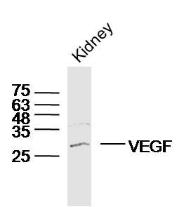
[IF=6.832] Liu, Jingyu. et al. Therapeutic effect of adipose stromal vascular fraction spheroids for partial bladder outlet obstruction induced underactive bladder. Stem Cell Res Ther. 2022 Dec;13(1):1-16 WB ; Rat.
[IF=3.528] Jie Wang. et al. A radiomics model based on DCE-MRI and DWI may improve the prediction of estimating IDH1 mutation and angiogenesis in gliomas. Eur J Radiol. 2022 Feb;147:110141 IHC ; Human.
[IF=9.933] Qiuyue Ding. et al. Bioinspired Multifunctional Black Phosphorus Hydrogel with Antibacterial and Antioxidant Properties: A Stepwise Countermeasure for Diabetic Skin Wound Healing. 2022 Feb 19 WB ; Human.
[IF=9.078] Lin Zou. et al. Icariin-releasing 3D printed scaffold for bone regeneration. Compos Part B-Eng. 2022 Mar;232:109625 IHC ; Rat.
[IF=3.411] Li Zhi-xin. et al. circGLI3 Inhibits Oxidative Stress by Regulating the miR-339-5p/VEGFA Axis in IPESLCJ2 Cells. Biomed Res Int. 2021;2021:1086206 WB ; Pig.
[IF=4.268] Jian-xia Wen. et al. Therapeutic Effects and Potential Mechanism of Dehydroevodiamine on N-Methyl-N′-Nitro-N-Nitrosoguanidine-Induced Chronic Atrophic Gastritis. Phytomedicine. 2021 Jun;:153619 WB,IHC ; Rat.
[IF=7.242] Yong Tang. et al. Laminin alpha 4 promotes bone regeneration by facilitating cell adhesion and vascularization. Acta Biomater. 2021 Mar;: WB,IF,IHC ; Mouse.
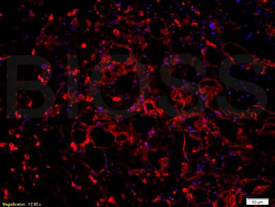
[IF=3.216] Yanhong Wang. et al. Intermedin Alleviates Renal Ischemia-Reperfusion Injury and Enhances Neovascularization in Wistar Rats. Drug Des Dev Ther. 2020; 14: 4825–4834 IHC ; Rat.
[IF=2.283] Ismet Hortu. et al. Oxytocin and cabergoline alleviate ovarian hyperstimulation syndrome (OHSS) by suppressing vascular endothelial growth factor (VEGF) in an experimental model. 2020 Nov 02 IHC ; Rat.
[IF=4.848] Hu Y et al. Non-Invasive Estimation of Glioma IDH1 Mutation and VEGF Expression by Histogram Analysis of Dynamic Contrast-Enhanced MRIFront Oncol.2020 Dec 8;10:593102. IHC ; Human.
[IF=4.784] Ding J et al. Anti-angiogenic effect of a chemically sulfated polysaccharide from Phellinus ribis by inhibiting VEGF/VEGFR pathway. Int J Biol Macromol. 2020 Mar 11;154:72-81. IHC,WB ; mouse.
[IF=2.705] Zhang Q et al. The circular RNA hsa_circ_0007623 acts as a sponge of microRNA-297 and promotes cardiac repair. Biochem Biophys Res Commun. 2020 Mar 19;523(4):993-1000. IHSLCP ; Mouse.
[IF=2.479] Wang H et al. The effects of a hirudin/liposome complex on a diabetic nephropathy rat model. BMC Complement Altern Med. 2019 Jun 6;19(1):118. IHSLCP ; Rat.
[IF=1.694] Yahuafai J et al. Anticancer Efficacy of the Combination of Berberine and PEGylated Liposomal Doxorubicin in Meth A Sarcoma-Bearing Mice.Biol Pharm Bull. 2018;41(7):1103-1106. WB ; Human.
[IF=3.349] Bi SX et al. The antitumour growth and antiangiogenesis effects of xanthatin in murine glioma dynamically evaluated by dynamic contrast‐enhanced magnetic resonance imaging.(2018) Phytother Res. Oct 22. IHC ; Rat、Mouse.
[IF=2.159] Liu X et al. The impact of ALDH2 activation by Alda-1 on the expression of VEGF in the hippocampus of a rat model of post-MI depression. Neurosci Lett. 2018 May 1;674:156-161. WB&IHC ; Rat.
[IF=0] Ren et al. Interferon-γ and celecoxib inhibit lung-tumor growth through modulating M2/M1 macrophage ratio in the tumor microenvironment. (2014) Drug.Des.Devel.Ther. 8:1527-38 IHC ; Mouse.
[IF=1.075] Yao, Qian, Yingxia Zhou, and Bing Li. "Thymoquinone (TQ) inhibits corneal neovascularization in a rabbit model." Int J Clin Exp Med 9.8 (2016): 15907-15913. IHSLCP ; Rabbit.
[IF=1.922] Chi et al. Transdermal estrogen gel and oral aspirin combination therapy improves fertility prognosis via the promotion of endometrial receptivity in moderate to severe intrauterine adhesion. (2018) Mol.Med.Rep. 17:6337-6344 IHC ; Human.
[IF=2.1] Zhenjie Huang. et al. Protective effects of different anti‑inflammatory drugs on tracheal stenosis following injury and potential mechanisms. Mol Med Rep. 2021 May;23(5):1-11 WB ; Rabbit.
[IF=6.291] Peng Zheng. et al. Alleviative effect of melatonin on the decrease of uterine receptivity caused by blood ammonia through ROS/NF-κB pathway in dairy cow. Ecotox Environ Safe. 2022 Feb;231:113166 ELISA ; Bovine.
[IF=5.923] Liang Hu. et al. Transient Activation of Hedgehog Signaling Inhibits Cellular Senescence and Inflammation in Radiated Swine Salivary Glands through Preserving Resident Macrophages. Int J Mol Sci. 2021 Jan;22(24):13493 WB,IHC ; Pig.
[IF=4.064] Ming Ni. et al. Sox11 Modified Tendon-Derived Stem Cells Promote the Repair of Osteonecrosis of Femoral Head:. Cell Transplant. 2021;(): WB ; Rabbit.
[IF=6.684] Huihua Kai. et al. LncRNA NORAD Promotes Vascular Endothelial Cell Injury and Atherosclerosis Through Suppressing VEGF Gene Transcription via Enhancing H3K9 Deacetylation by Recruiting HDAC6. Front Cell Dev Biol. 2021; 9: 701628 WB ; Human.
[IF=3.375] Shun Zhang. et al. PPARγ induces the paroxysm of endometriosis by regulating the transcription of MAT2A gene. Am J Transl Res. 2021; 13(3): 1377–1388 IHC ; Human.
[IF=2.73] Ling Zhou. et al. Psoriatic mesenchymal stem cells stimulate the angiogenesis of human umbilical vein endothelial cells in vitro. Microvasc Res. 2021 Jul;136:104151 WB ; Human.
[IF=5.201] Qiong Li. et al. CD73+ Mesenchymal Stem Cells Ameliorate Myocardial Infarction by Promoting Angiogenesis. Front Cell Dev Biol. 2021; 9: 637239 WB,IHC ; Rat.
[IF=10.652] Pei Liu. et al. Biphasic CK2.1-coated β-glycerophosphate chitosan/LL37-modified layered double hydroxide chitosan composite scaffolds enhance coordinated hyaline cartilage and subchondral bone regeneration. Chem Eng J. 2021 Aug;418:129531 IHC ; Rabbit.
[IF=4.197] Xuewen Qian. et al. Acidosis induces synovial fibroblasts to release vascular endothelial growth factor via acid-sensitive ion channel 1a. Lab Invest. 2020 Aug;101(3):280-291 WB,IHC ; Human.
[IF=3.375] Wei P. et al. Nintedanib ameliorates tracheal stenosis by activating HDAC2 and suppressing IL-8 and VEGF in rabbit.. Am J Transl Res. 2020 Aug;12(8):4739-4748 WB ; Rabbit.
[IF=2.055] Li-Hua Muet al. Antiangiogenic effects of AG36, a triterpenoid saponin from Ardisia gigantifolia stapf. J Nat Med
. 2020 Sep;74(4):732-740. WB ; Human.
[IF=1.918] Mao XB et al. Sirtuin (Sirt) 3 Overexpression Prevents Retinopathy in Streptozotocin-Induced Diabetic Rats. Med Sci Monit
. 2020 May 27;26:e920883. WB ; rat.
[IF=1.2] Özel F et al. Protective effect of alpha lipoic acid on 4-vinylcyclohexene diepoxide induced primary ovarian failure in female rats. Taiwan J Obstet Gynecol. 2020 Mar;59(2):293-300. IHC ; rat.
[IF=3.973] Liu N et al. Polyphotosensitizer Nanogels for GSH-Responsive Histone Deacetylase Inhibitors delivery and enhanced Cancer Photodynamic Therapy. Colloids Surf B Biointerfaces. 2019 Dec 23;188:110753. IHSLCP ; Mouse.
[IF=3.44] Han Y et al. Studies on bacterial cellulose/poly vinyl alcohol (BC/PVA) hydrogel composites as tissue-engineered corneal stroma. Biomed Mater. 2019 Nov 12. IHSLCP ; Rabbit.
[IF=2.807] Ma WJ et al. Biliary antibiotics irrigation for E. coli-induced chronic proliferative cholangitis and hepatolithiasis: A pathophysiological study in rabbits. Clin Res Hepatol Gastroenterol. 2019 Aug 13. pii: S2210-7401(19)30173-1. WB ; Rabbit.
[IF=3.103] Li R et al. Lithium chloride promoted hematoma resolution after intracerebral hemorrhage through GSK-3β-mediated pathways-dependent microglia phagocytosis and M2-phenotype differentiation, angiogenesis and neurogenesis in a rat model. Brain Res Bull. 2019 Jul 17;152:117-127. WB ; Rat.
[IF=1.872] Li Z et al. Angiogenesis and bone regeneration by allogeneic mesenchymal stem cell intravenous transplantation in rabbit model of avascular necrotic femoral head. J Surg Res. 2013 Jul;183(1):193-203. IHSLCP ; Rabbit.
[IF=3.115] Zhen-zhenChen et al. Bu Yang Huan Wu Decoction prevents reperfusion injury following ischemic stroke in rats via inhibition of HIF-1 α, VEGF and promotion β-ENaC expression (2018)Journal of Ethnopharmacology Sep 13;228:70-81. WB ; Rat.
[IF=1.766] Yuan X et al. Significance of nuclear magnetic resonance combined with Ki-67 and VEGF detection in the diagnosis and prognosis evaluation of brain glioma.J BUON. 2018 Mar-Apr;23(2):410-415. IHSLCP ; Human.
[IF=0] Gautam et al. Implantation of autologous adipose-derived cells reconstructs functional urethral sphincters in rabbit cryoinjured urethra. (2014) Tissue.Eng.Part.. 20:1971-9 IHSLCP ; Rabbit.
[IF=1.69] Li et al. Curcumin attenuates the development of thoracic aortic aneurysm by inhibiting VEGF expression and inflammation. (2017) Mol.Med.Rep. 16:4455-4462 IHSLCP, WB ; Human, Rat.