[IF=4.73] Erdal Ayhan Işik. et al. Use of Erythropoietin and Fibrin Glue Mixture for Peripheral Nerve Repair. Plast Reconstr Surg. 2022 Feb;149(2):395-403 IHC ; Rat.
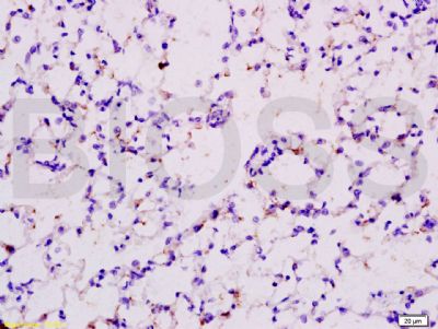
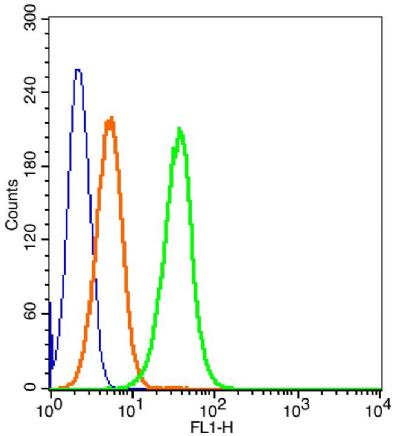
[IF=7.561] Wen Zhang. et al. Extracellular CIRP-Impaired Rab26 Restrains EPOR-Mediated Macrophage Polarization in Acute Lung Injury. Front Immunol. 2021; 12: 768435 FC ; Mouse.
[IF=3.414] Li ZH et al. You-Gui-Yin improved the reproductive dysfunction of male rats with chronic kidney disease via regulating the HIF1α-STAT5 pathway. J Ethnopharmacol. 2020 Jan 10;246:11248. WB&IHSLCP ; Rat.
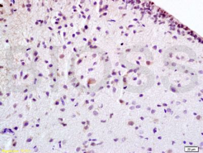
[IF=1.56] Zhong, Cheng, and Xueyuan Zhang. "Age‑associated expression of erythropoietin and its receptor in rat spiral ganglion neurons and its association with neuronal apoptosis and hearing alterations." Molecular Medicine Reports. IHSLCP ; Rat.
[IF=3.35] Li, Xu, et al. "Oxidative stress induces the decline of brain EPO expression in aging rats." Experimental Gerontology (2016). WB ; Rat.
[IF=3.414] Tang Q et al. Aqueous extract from You-Gui-Yin ameliorates cognitive impairment of chronic renal failure micethrough targeting hippocampal CaMKIIα/CREB/BDNF and EPO/EPOR pathways. J Ethnopharmacol. 2019 Jul 15;239:111925. WB ; Mouse.
[IF=12.479] Tao Liu. et al. Multifaceted roles of a bioengineered nanoreactor in repressing radiation-induced lung injury. Biomaterials. 2021 Oct;277:121103 IF ; Human.
[IF=2.583] Li YJ et al. Vasculogenic Mimicry Formation Is Associated with Erythropoietin Expression but Not with Erythropoietin Receptor Expression in Cervical Squamous Cell Carcinoma. BioMed Research International, 2019. IHSLCP ; Human.
[IF=2.78] Zhong et al. Protective effect of adenovirus-mediated erythropoietin expression on the spiral ganglion neurons in the rat inner ear. (2018) Int.J.Mol.Med. 41:2669-2677 IHC ; Human.