The PAF receptor binds platelet-activating factor (PAF) and is thought to mediate its action via a G protein that activates a phosphatidylinositol-calcium second messenger system. PAF is a chemotactic phospholipid mediator that possesses potent inflammatory, smooth-muscle contractile and hypotensive activity. It has been implicated as a mediator in diverse pathologic processes, such as allergy, asthma, septic shock, arterial thrombosis, and inflammatory processes.
The PAF receptor is induced by granulocyte macrophage colony-stimulating factor (GM-CSF), interleukin-5 and n-butyrate. A diverse range of compounds act as PAF receptor antagonists; these may be useful pharmacologically.
Function:
Receptor for platelet activating factor, a chemotactic phospholipid mediator that possesses potent inflammatory, smooth-muscle contractile and hypotensive activity. Seems to mediate its action via a G protein that activates a phosphatidylinositol-calcium second messenger system.
Subunit:
Interacts with ARRB1.
Subcellular Location:
Cell membrane; Multi-pass membrane protein.
Tissue Specificity:
Expressed in the placenta, lung, left and right heart ventricles, heart atrium, leukocytes and differentiated HL-60 granulocytes.
Similarity:
Belongs to the G-protein coupled receptor 1 family.
SWISS:
P25105
Gene ID:
5724
Database links:
Entrez Gene: 5724 Human
Entrez Gene: 19204 Mouse
Entrez Gene: 58949 Rat
Omim: 173393 Human
SwissProt: P25105 Human
SwissProt: Q62035 Mouse
SwissProt: Q9XSD4 Pig
SwissProt: P9202 Rat
SwissProt: P35366 Rhesus monkey
Unigene: 433540 Human
Unigene: 725944 Human
Unigene: 89389 Mouse
Unigene: 10137 Rat
| Picture |
Sample:
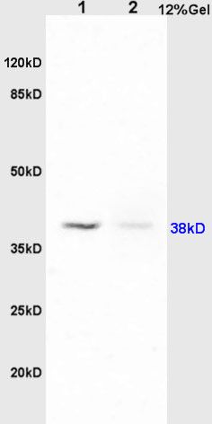
Lane1: Lung(Rat) Lysate at 30 ug
Lane2: Brain(Rat) Lysate at 30 ug
Primary: Anti-PTAFR (SL1478R) at 1:200 dilution;
Secondary: HRP conjugated Goat Anti-Rabbit IgG(SL0295G-HRP) at 1: 3000 dilution;
Predicted band size : 38kD
Observed band size : 38kD
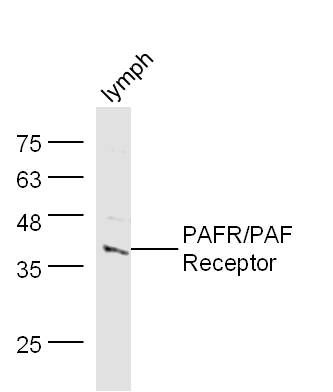
Sample: Lymph (Mouse) Lysate at 30 ug
Primary: Anti- RAFR/PAF (SL1478R) at 1/300 dilution
Secondary: IRDye800CW Goat Anti-Rabbit IgG at 1/10000 dilution
Predicted band size: 38 kD
Observed band size: 38 kD
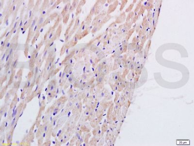
Tissue/cell: rat heart tissue; 4% Paraformaldehyde-fixed and paraffin-embedded;
Antigen retrieval: citrate buffer ( 0.01M, pH 6.0 ), Boiling bathing for 15min; Block endogenous peroxidase by 3% Hydrogen peroxide for 30min; Blocking buffer (normal goat serum,SLC0005) at 37℃ for 20 min;
Incubation: Anti-PTAFR Polyclonal Antibody, Unconjugated(SL1478R) 1:200, overnight at 4°C, followed by conjugation to the secondary antibody(SP-0023) and DAB(SLC0010) staining
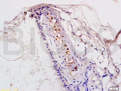
Tissue/cell: rat lung tissue; 4% Paraformaldehyde-fixed and paraffin-embedded;
Antigen retrieval: citrate buffer ( 0.01M, pH 6.0 ), Boiling bathing for 15min; Block endogenous peroxidase by 3% Hydrogen peroxide for 30min; Blocking buffer (normal goat serum,SLC0005) at 37℃ for 20 min;
Incubation: Anti-PTAFR Polyclonal Antibody, Unconjugated(SL1478R) 1:200, overnight at 4°C, followed by conjugation to the secondary antibody(SP-0023) and DAB(SLC0010) staining
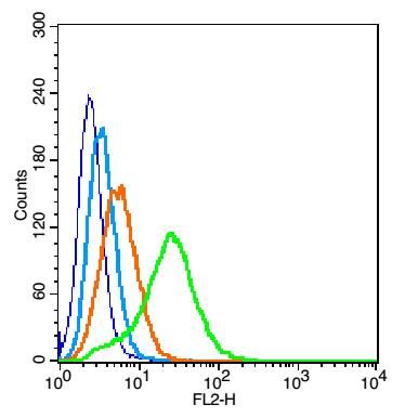
Blank control: 293T cells(blue).
Primary Antibody:Rabbit Anti-PAFR/PAF antibody(SL1478R), Dilution: 1μg in 100 μL 1X PBS containing 0.5% BSA;
Isotype Control Antibody: Rabbit IgG(orange) ,used under the same conditions );
Secondary Antibody: Goat anti-rabbit IgG-PE(white blue), Dilution: 1:200 in 1 X PBS containing 0.5% BSA.
Protocol
The cells were fixed with 2% paraformaldehyde (10 min). Primary antibody (SL1478R, 1μg /1x10^6 cells) were incubated for 30 min on the ice, followed by 1 X PBS containing 0.5% BSA + 1 0% goat serum (15 min) to block non-specific protein-protein interactions. Then the Goat Anti-rabbit IgG/PE antibody was added into the blocking buffer mentioned above to react with the primary antibody at 1/200 dilution for 30 min on ice. Acquisition of 20,000 events was performed.
|
|
|