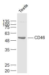
Sample:
Testis (Mouse) Lysate at 40 ug
Primary: Anti-CD46 (SL1529R) at 1/1000 dilution
Secondary: IRDye800CW Goat Anti-Rabbit IgG at 1/20000 dilution
Predicted band size: 43 kD
Observed band size: 55 kD
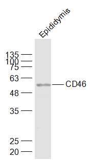
Sample:
Epididymis (Mouse) Lysate at 40 ug
Primary: Anti-CD46 (SL1529R) at 1/1000 dilution
Secondary: IRDye800CW Goat Anti-Rabbit IgG at 1/20000 dilution
Predicted band size: 43 kD
Observed band size: 56 kD
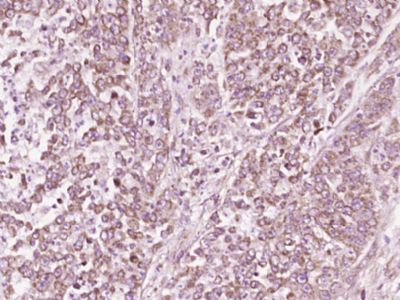
Paraformaldehyde-fixed, paraffin embedded (Human colon carcinoma); Antigen retrieval by boiling in sodium citrate buffer (pH6.0) for 15min; Block endogenous peroxidase by 3% hydrogen peroxide for 20 minutes; Blocking buffer (normal goat serum) at 37°C for 30min; Antibody incubation with (CD46) Polyclonal Antibody, Unconjugated (SL1529R) at 1:400 overnight at 4°C, followed by operating according to SP Kit(Rabbit) (sp-0023) instructionsand DAB staining.
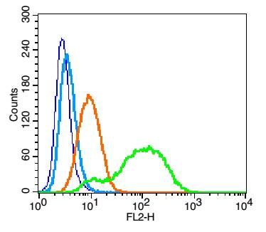
Blank control: U937 (blue).
Primary Antibody: Rabbit Anti- CD46 antibody(SL1529R), Dilution: 1μg in 100 μL 1X PBS containing 0.5% BSA;
Isotype Control Antibody: Rabbit IgG(orange) ,used under the same conditions );
Secondary Antibody: Goat anti-rabbit IgG-PE(white blue), Dilution: 1:200 in 1 X PBS containing 0.5% BSA.
Protocol
The cells were fixed with 2% paraformaldehyde (10 min). Primary antibody (SL1529R, 1μg /1x10^6 cells) were incubated for 30 min on the ice, followed by 1 X PBS containing 0.5% BSA + 1 0% goat serum (15 min) to block non-specific protein-protein interactions. Then the Goat Anti-rabbit IgG/PE antibody was added into the blocking buffer mentioned above to react with the primary antibody at 1/200 dilution for 30 min on ice. Acquisition of 20,000 events was performed.
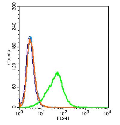
Blank control: 293T(blue).
Primary Antibody:Rabbit Anti-CD46 antibody(SL1529R), Dilution: 1μg in 100 1μL 1X PBS containing 0.5% BSA(green);
Isotype Control Antibody: Rabbit IgG(orange), used under the same conditions );
Secondary Antibody: Goat anti-rabbit IgG-PE(white blue), Dilution: 1:200 in 1 X PBS containing 0.5% BSA.
protocol
The cells were washed twice with phosphate-buffered saline (PBS).The cells were then incubated in 1 X PBS containing 0.5% BSA + 10% goat serum (15 min) to block non-specific protein-protein interactions followed by the antibody (SL1529R, 1μg/1x10^6 cells) for 30 min on ice. The secondary antibody used was Goat Anti-rabbit IgG/PE antibody at 1/200 dilution for 30 min on ice.Acquisition of 20,000 events was performed.
|