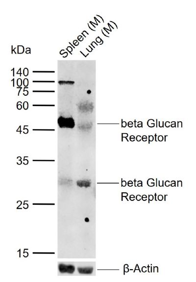
Sample:
Lane 1: Mouse Spleen tissue lysates
Lane 2: Mouse Lung tissue lysates
Primary: Anti-beta Glucan Receptor (SL2455R) at 1/1000 dilution
Secondary: IRDye800CW Goat Anti-Rabbit IgG at 1/20000 dilution
Predicted band size: 28 kDa
Observed band size: 28,47 kDa
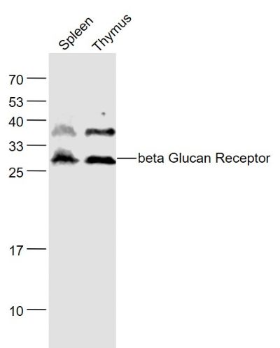
Sample:
Spleen (Mouse) Lysate at 40 ug
Thymus (Mouse) Lysate at 40 ug
Primary: Anti- beta Glucan Receptor (SL2455R) at 1/1000 dilution
Secondary: IRDye800CW Goat Anti-Rabbit IgG at 1/20000 dilution
Predicted band size: 28 kD
Observed band size: 28 kD
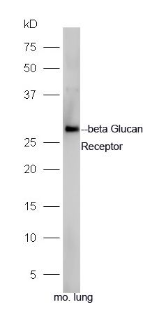
Sample: Lung(Mouse) lysate at 30ug;
Primary: Anti-beta Glucan Receptor (SL2455R) at 1:300 dilution;
Secondary: HRP conjugated Goat-Anti-rabbit IgG(SL0295G-HRP) at 1:5000 dilution;
Predicted band size: 28 kD
Observed band size: 28 kD
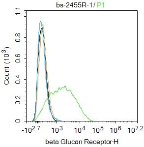
Blank control:RAW264.7.
Primary Antibody (green line): Rabbit Anti-Iba1 antibody (SL2455R)
Dilution: 1ug/Test;
Secondary Antibody (white blue line) : Goat anti-Rabbit IgG-AF488
Dilution: 0.5ug/Test.
Isotype control(orange line):Normal Rabbit IgG
Protocol
The cells were incubated in 5%BSA to block non-specific protein-protein interactions for 30 min at room temperature .Cells stained with Primary Antibody for 30 min at room temperature. The secondary antibody used for 40 min at room temperature. Acquisition of 20,000 events was performed.
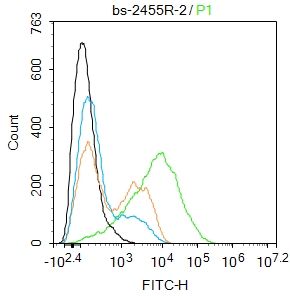
Blank control:THP-1.
Primary Antibody (green line): Rabbit Anti-beta Glucan Receptor antibody (SL2455R)
Dilution: 2μg /10^6 cells;
Isotype Control Antibody (orange line): Rabbit IgG .
Secondary Antibody : Goat anti-rabbit IgG-FITC
Dilution: 1μg /test.
Protocol
The cells were incubated in 5%BSA to block non-specific protein-protein interactions for 30 min at room temperature .Cells stained with Primary Antibody for 30 min at room temperature. The secondary antibody used for 40 min at room temperature. Acquisition of 20,000 events was performed.
|