Histones are basic nuclear proteins that are responsible for the nucleosome structure of the chromosomal fiber in eukaryotes. Nucleosomes consist of approximately 146 bp of DNA wrapped around a histone octamer composed of pairs of each of the four core histones (H2A, H2B, H3, and H4). The chromatin fiber is further compacted through the interaction of a linker histone, H1, with the DNA between the nucleosomes to form higher order chromatin structures. This gene is intronless and encodes a replication-dependent histone that is a member of the histone H3 family. Transcripts from this gene lack polyA tails; instead, they contain a palindromic termination element. This gene is located separately from the other H3 genes that are in the histone gene cluster on chromosome 6p22-p21.3. [provided by RefSeq, Aug 2015]
Function:
Core component of nucleosome. Nucleosomes wrap and compact DNA into chromatin, limiting DNA accessibility to the cellular machineries which require DNA as a template. Histones thereby play a central role in transcription regulation, DNA repair, DNA replication and chromosomal stability. DNA accessibility is regulated via a complex set of post-translational modifications of histones, also called histone code, and nucleosome remodeling. H3 is deposited into chromatin exclusively through a DNA replication-coupled pathway that can be associated with either DNA duplication or DNA repair synthesis during meiotic homologous recombination.
Subunit:
The nucleosome is a histone octamer containing two molecules each of H2A, H2B, H3 and H4 assembled in one H3-H4 heterotetramer and two H2A-H2B heterodimers. The octamer wraps approximately 147 bp of DNA. Interacts with GCN5, whereby H3S10ph increases histone-protein interactions. Interacts with PDD1 and PDD3.
Subcellular Location:
Nucleus. Chromosome. Note=Localizes to both the large, transcriptionally active, somatic macronucleus (MAC) and the small, transcriptionally inert, germ line micronucleus (MIC).
Post-translational modifications:
Phosphorylated to form H3S10ph. H3S10ph promotes subsequent H3K14ac formation by GCN5. H3S10ph is only found in the mitotically dividing MIC, but not in the amitotically dividing MAC. H3S10ph is correlated with chromosome condensation during mitotic or meiotic micronuclear divisions.
Acetylation of histone H3 leads to transcriptional activation. H3K14ac formation by GCN5 is promoted by H3S10ph. H3K9acK14ac is the preferred acetylated form of newly synthesized H3. Acetylation occurs almost exclusively in the MAC.
Methylated to form H3K4me. H3K4me is only found in the transcriptionally active MAC. Methylated to form H3K9me in developing MACs during conjugation, when genome-wide DNA elimination occurs. At this stage, H3K9me specifically occurs on DNA sequences being eliminated (IES), probably targeted by small scan RNAs (scnRNAs) bound to IES, and is required for efficient IES elimination. H3K9me is required for the interaction with the chromodomains of PDD1 and PDD3.
The full-length protein H3S (slow migrating) is converted to H3F (fast migrating) by proteolytic removal of the first 6 residues. H3F is unique to MIC, and processing seems to occur regularly each generation at a specific point in the cell cycle.
Similarity:
Belongs to the histone H3 family.
SWISS:
P68431
Gene ID:
8350
Database links:
Entrez Gene: 8350 Human
Entrez Gene: 8351 Human
Entrez Gene: 8352 Human
Entrez Gene: 8353 Human
Entrez Gene: 8354 Human
Entrez Gene: 8355 Human
Entrez Gene: 8356 Human
Entrez Gene: 8357 Human
Entrez Gene: 8358 Human
Entrez Gene: 8968 Human
Entrez Gene: 260423 Mouse
Entrez Gene: 319148 Mouse
Entrez Gene: 319149 Mouse
Entrez Gene: 319150 Mouse
Entrez Gene: 319151 Mouse
Entrez Gene: 319152 Mouse
Entrez Gene: 319153 Mouse
Entrez Gene: 72198 Mouse
Entrez Gene: 97908 Mouse
Entrez Gene: 100364501 Rat
Entrez Gene: 100365669 Rat
Entrez Gene: 291159 Rat
Entrez Gene: 314977 Rat
Entrez Gene: 364716 Rat
Entrez Gene: 679950 Rat
Entrez Gene: 679994 Rat
Entrez Gene: 136511 Rat
Entrez Gene: 136599 Rat
Entrez Gene: 682330 Rat
Entrez Gene: 691496 Rat
SwissProt: P68431 Human
SwissProt: P84243 Human
SwissProt: Q16695 Human
SwissProt: Q6NXT2 Human
SwissProt: Q71DI3 Human
SwissProt: P68433 Mouse
SwissProt: P84228 Mouse
SwissProt: Q6LED0 Rat
组蛋白的基因非常保守,在亲缘关系较远的种属中,四种组蛋白(H2A、H2A、H3、H4)氨基酸序列都非常相似,如海胆组织H3的氨基酸序列与来自小牛胸腺的H3的氨基酸序列间只有一个氨基酸的差异,小牛胸腺的H3的氨基酸序列与豌豆的H3也很相似。组蛋白是细胞核内的一种碱性核蛋白,抗组蛋白抗体即是以组蛋白为靶抗原的一种自身,是抗核抗体的一种。分子量:16-18KDa。主要与药物性红斑狼疮、系统性红斑狼疮、类风湿关节炎有关。
| Picture |
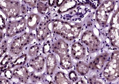
Paraformaldehyde-fixed, paraffin embedded (Human kidney); Antigen retrieval by boiling in sodium citrate buffer (pH6.0) for 15min; Block endogenous peroxidase by 3% hydrogen peroxide for 20 minutes; Blocking buffer (normal goat serum) at 37°C for 30min; Antibody incubation with (Phospho-Histone H3 (Ser10)) Polyclonal Antibody, Unconjugated (SL3186R) at 1:400 overnight at 4°C, followed by operating according to SP Kit(Rabbit) (sp-0023) instructionsand DAB staining.
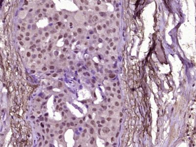
Paraformaldehyde-fixed, paraffin embedded (Human breast carcinoma); Antigen retrieval by boiling in sodium citrate buffer (pH6.0) for 15min; Block endogenous peroxidase by 3% hydrogen peroxide for 20 minutes; Blocking buffer (normal goat serum) at 37°C for 30min; Antibody incubation with (Phospho-Histone H3 (Ser10)) Polyclonal Antibody, Unconjugated (SL3186R) at 1:400 overnight at 4°C, followed by operating according to SP Kit(Rabbit) (sp-0023) instructionsand DAB staining.
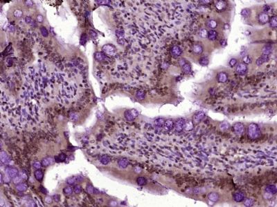
Paraformaldehyde-fixed, paraffin embedded (Rat colon); Antigen retrieval by boiling in sodium citrate buffer (pH6.0) for 15min; Block endogenous peroxidase by 3% hydrogen peroxide for 20 minutes; Blocking buffer (normal goat serum) at 37°C for 30min; Antibody incubation with (Phospho-Histone H3 (Ser10)) Polyclonal Antibody, Unconjugated (SL3186R) at 1:400 overnight at 4°C, followed by operating according to SP Kit(Rabbit) (sp-0023) instructionsand DAB staining.
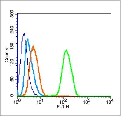
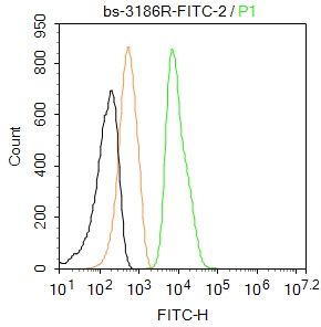
Blank control (blue line): Hela(fixed with 70% ethanol (Overnight at 4℃) and then permeabilized with 90% ice-cold methanol for 30 min on ice).
Primary Antibody (green line): Rabbit Anti-Phospho-Histone H3 (Ser10) antibody (SL3186R),Dilution: 0.2μg /10^6 cells;
Isotype Control Antibody (orange line): Rabbit IgG .
Secondary Antibody (white blue line): Goat anti-rabbit IgG-FITC,Dilution: 1μg /test.
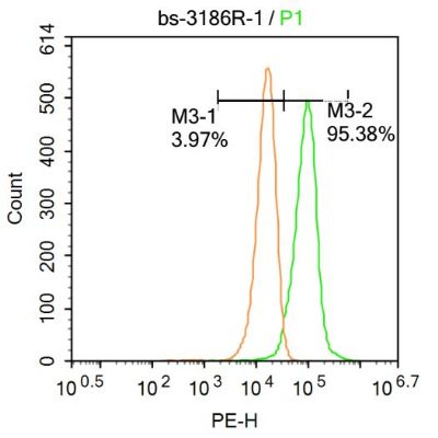
Blank control:Hela.
Primary Antibody (green line): Rabbit Anti-Histone H3 (Di Methyl K36) antibody (SL3186R-FITC)
Dilution: 2μg /10^6 cells;
Isotype Control Antibody (orange line): Rabbit IgG .
Protocol
The cells were fixed with 4% PFA (10min at room temperature)and then permeabilized with 90% ice-cold methanol for 20 min at -20℃. The cells were then incubated in 5%BSA to block non-specific protein-protein interactions for 30 min at room temperature .Cells stained with Primary Antibody for 30 min at room temperature. Acquisition of 20,000 events was performed.
Blank control:U-87MG.
Primary Antibody (green line): Rabbit Anti-Phospho-Histone H3 antibody (SL3186R)
Dilution: 1μg /10^6 cells;
Isotype Control Antibody (orange line): Rabbit IgG .
Secondary Antibody : Goat anti-rabbit IgG-AF647
Dilution: 1μg /test.
Protocol
The cells were fixed with 4% PFA (10min at room temperature)and then permeabilized with 90% ice-cold methanol for 20 min at-20℃. The cells were then incubated in 5%BSA to block non-specific protein-protein interactions for 30 min at at room temperature .Cells stained with Primary Antibody for 30 min at room temperature. The secondary antibody used for 40 min at room temperature. Acquisition of 20,000 events was performed.
|
|
|