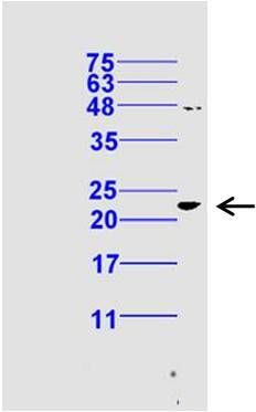
Sample: Testis (Mouse) Tissue Lysate at 30 ug
Primary: Anti-phospho-Bax (Ser184) (SL3010R)at 1/300 dilution
Secondary: IRDye800CW Goat Anti-Rabbit IgG at 1/20000 dilution
Predicted band size: 21 kD
Observed band size: 22 kD
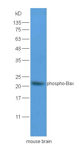
Sample:Brain(Mouse) lysate at 30ug;
Primary: Anti-phospho-Bax (Ser184) (SL3010R) at 1:200 dilution;
Secondary: HRP conjugated Goat Anti-Rabbit IgG(SL0295G-HRP) at 1: 5000 dilution;
Predicted band size : 21kD
Observed band size : 21kD
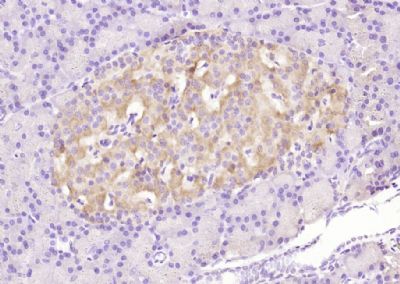
Paraformaldehyde-fixed, paraffin embedded (rat pancreas); Antigen retrieval by boiling in sodium citrate buffer (pH6.0) for 15min; Block endogenous peroxidase by 3% hydrogen peroxide for 20 minutes; Blocking buffer (normal goat serum) at 37°C for 30min; Antibody incubation with (phospho-Bax (Ser184)) Polyclonal Antibody, Unconjugated (SL3010R) at 1:200 overnight at 4°C, followed by operating according to SP Kit(Rabbit) (sp-0023) instructionsand DAB staining.
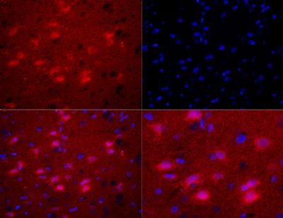
Tissue/cell: rat brain tissue;4% Paraformaldehyde-fixed and paraffin-embedded;
Antigen retrieval: citrate buffer ( 0.01M, pH 6.0 ), Boiling bathing for 15min; Blocking buffer (normal goat serum,SLC0005) at 37℃ for 20 min;
Incubation: Anti-phospho-Bax(Ser184) Polyclonal Antibody, Unconjugated(SL3010R) 1:200, overnight at 4°C; The secondary antibody was Goat Anti-Rabbit IgG, Cy3 conjugated (SL0295G-Cy3)used at 1:200 dilution for 40 minutes at 37°C. DAPI(5ug/ml,blue,SLC0033) was used to stain the cell nuclei
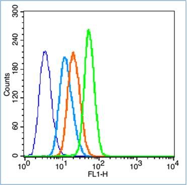
Blank control (blue line): HL60 (fixed with 2% paraformaldehyde (10 min) and then permeabilized with 0.1% PBS-Tween for 20 min at room temperature).
Primary Antibody (green line): Rabbit Anti-phospho-Bax (Ser184) antibody (SL3010R),Dilution: 3μg /10^6 cells;
Isotype Control Antibody (orange line): Rabbit IgG .
Secondary Antibody (white blue line): Goat anti-rabbit IgG-FITC,Dilution: 1μg /test.
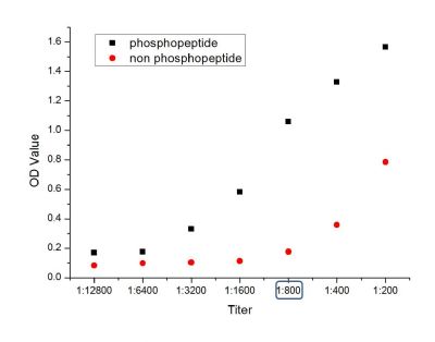
phosphopeptide
non phosphopeptide
|