Home > Product > Antibody > Rabbit Anti-Phospho-Smad1 (Ser206) antibody
Smad1 (phospho S206); Smad1 (phospho Ser206); p-Smad1 (Ser206); Mothers against decapentaplegic homolog 1; BSP 1; BSP1; BSP-1; Dwarfin A; DwfA; hSMAD 1; hSMAD1; JV 41; JV4 1; JV4-1; JV41; MAD mothers against decapentaplegic homolog 1; Mad related protein
Cat:
SL3417R
Species Reactivity:
Human,Mouse,(predicted: Rat,)
Immunogen:
KLH conjugated Synthesised phosphopeptide derived from human Smad1 around the phosphorylation site of Ser206:PH(p-S)PT
Format:
Liquid
Storage instructions:
Shipped at 4℃. Store at -20 °C for one year. Avoid repeated freeze/thaw cycles.
Concentration:
1mg/ml
Clonality:
Polyclonal
Isotype:
IgG
Applications:
WB=1:500-2000ELISA=1:5000-10000IHC-P=1:100-500IHC-F=1:100-500ICC=1:100-500IF=1:100-500(Paraffin sections need to do antigen repair)not yet tested in other applications.optimal dilutions/concentrations should be determined by the end user.
Host:
Rabbit
Product Overview:
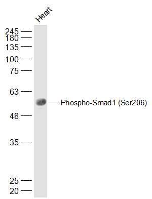
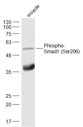
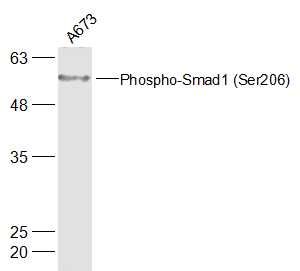
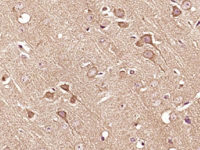
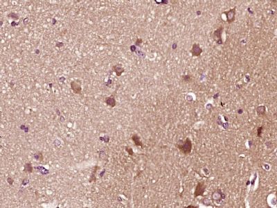
Sample: Heart (Mouse) Lysate at 40 ugPrimary: Anti-Phospho-Smad1 (Ser206) (SL3417R) at 1/500 dilutionSecondary: IRDye800CW Goat Anti-Rabbit IgG at 1/20000 dilutionPredicted band size: 52 kDObserved band size: 52 kDSample: Muscle(Mouse) Cell Lysate at 40 ugPrimary: Anti-Phospho-Smad1 (Ser206) (SL3417R) at 1/300 dilutionSecondary: IRDye800CW Goat Anti-Rabbit IgG at 1/20000 dilutionPredicted band size: 52 kDObserved band size: 52 kDSample: A673(Human) Cell Lysate at 30 ugPrimary: Anti-Phospho-Smad1 (Ser206) (SL3417R) at 1/500 dilutionSecondary: IRDye800CW Goat Anti-Rabbit IgG at 1/20000 dilutionPredicted band size: 52 kDObserved band size: 52 kDParaformaldehyde-fixed, paraffin embedded (mouse brain tissue); Antigen retrieval by boiling in sodium citrate buffer (pH6.0) for 15min; Block endogenous peroxidase by 3% hydrogen peroxide for 20 minutes; Blocking buffer (normal goat serum) at 37°C for 30min; Antibody incubation with (Smad1 (Ser206)) Polyclonal Antibody, Unconjugated (SL3417R) at 1:400 overnight at 4°C, followed by operating according to SP Kit(Rabbit) (sp-0023) instructionsand DAB staining.Paraformaldehyde-fixed, paraffin embedded (human brain glioma); Antigen retrieval by boiling in sodium citrate buffer (pH6.0) for 15min; Block endogenous peroxidase by 3% hydrogen peroxide for 20 minutes; Blocking buffer (normal goat serum) at 37°C for 30min; Antibody incubation with (Smad1 (Ser206)) Polyclonal Antibody, Unconjugated (SL3417R) at 1:400 overnight at 4°C, followed by operating according to SP Kit(Rabbit) (sp-0023) instructionsand DAB staining.
More
Unit:
Price: $
Product PDFs
Datasheet:


The protein encoded by this gene belongs to the SMAD, a family of proteins similar to the gene products of the Drosophila gene 'mothers against decapentaplegic' (Mad) and the C. elegans gene Sma. SMAD proteins are signal transducers and transcriptional modulators that mediate multiple signaling pathways. This protein mediates the signals of the bone morphogenetic proteins (BMPs), which are involved in a range of biological activities including cell growth, apoptosis, morphogenesis, development and immune responses. In response to BMP ligands, this protein can be phosphorylated and activated by the BMP receptor kinase. The phosphorylated form of this protein forms a complex with SMAD4, which is important for its function in the transcription regulation. This protein is a target for SMAD-specific E3 ubiquitin ligases, such as SMURF1 and SMURF2, and undergoes ubiquitination and proteasome-mediated degradation. Alternatively spliced transcript variants encoding the same protein have been observed. [provided by RefSeq].

Function:
Transcriptional modulator activated by BMP (bone morphogenetic proteins) type 1 receptor kinase. SMAD1 is a receptor-regulated SMAD (R-SMAD). SMAD1/OAZ1/PSMB4 complex mediates the degradation of the CREBBP/EP300 repressor SNIP1.

Subunit:
Interacts with HGS, NANOG and ZCCHC12. May form trimers with another SMAD1 and the co-SMAD SMAD4. Interacts with PEBP2-alpha subunit, CREB-binding protein (CBP), p300, SMURF1, SMURF2 and HOXC8. Associates with ZNF423 or ZNF521 in response to BMP2 leading to activate transcription of BMP target genes. Interacts with SKOR1. Interacts (via MH2 domain) with LEMD3. Binding to LEMD3 results in at least a partial reduction of receptor-mediated phosphorylation. Forms a ternary complex with PSMB4 and OAZ1 before PSMB4 is incorporated into the 20S proteasome.

Subcellular Location:
Cytoplasm. Nucleus. Note=Cytoplasmic in the absence of ligand. Migrates to the nucleus when complexed with SMAD4. Co-localizes with LEMD3 at the nucleus inner membrane.

Tissue Specificity:
Ubiquitous. Highest expression seen in the heart and skeletal muscle.

Post-translational modifications:
Phosphorylated on serine by BMP type 1 receptor kinase.
Ubiquitin-mediated proteolysis by SMAD-specific E3 ubiquitin ligase SMURF1.

DISEASE:
Defects in SMAD1 may be a cause of primary pulmonary hypertension (PPH1) [MIM:178600]. A rare disorder characterized by plexiform lesions of proliferating endothelial cells in pulmonary arterioles. The lesions lead to elevated pulmonary arterial pression, right ventricular failure, and death. The disease can occur from infancy throughout life and it has a mean age at onset of 36 years. Penetrance is reduced. Although familial PPH1 is rare, cases secondary to known etiologies are more common and include those associated with the appetite-suppressant drugs.

Similarity:
Belongs to the dwarfin/SMAD family.
Contains 1 MH1 (MAD homology 1) domain.
Contains 1 MH2 (MAD homology 2) domain.

SWISS:
Q15797

Gene ID:
4086

Database links:

Entrez Gene: 4086 Human

Entrez Gene: 17125 Mouse

Entrez Gene: 25671 Rat

Omim: 601595 Human

SwissProt: Q15797 Human

SwissProt: P70340 Mouse

SwissProt: P97588 Rat

Unigene: 604588 Human

Unigene: 223717 Mouse

Unigene: 10635 Rat


Smads最新发现的TGF-β信号转导途径中一个重要的新基因家族.是TGF-β特异的细胞内信号转导分子,SMAD1属于受体激活的SMADs.有学者认为:Smad1在某些肿瘤中发生突变,有可能是一种肿瘤抑制基因。 Picture

Sample:
Heart (Mouse) Lysate at 40 ug
Primary: Anti-Phospho-Smad1 (Ser206) (SL3417R) at 1/500 dilution
Secondary: IRDye800CW Goat Anti-Rabbit IgG at 1/20000 dilution
Predicted band size: 52 kD
Observed band size: 52 kD
Sample:
Muscle(Mouse) Cell Lysate at 40 ug
Primary: Anti-Phospho-Smad1 (Ser206) (SL3417R) at 1/300 dilution
Secondary: IRDye800CW Goat Anti-Rabbit IgG at 1/20000 dilution
Predicted band size: 52 kD
Observed band size: 52 kD
Sample:
A673(Human) Cell Lysate at 30 ug
Primary: Anti-Phospho-Smad1 (Ser206) (SL3417R) at 1/500 dilution
Secondary: IRDye800CW Goat Anti-Rabbit IgG at 1/20000 dilution
Predicted band size: 52 kD
Observed band size: 52 kD
Paraformaldehyde-fixed, paraffin embedded (mouse brain tissue); Antigen retrieval by boiling in sodium citrate buffer (pH6.0) for 15min; Block endogenous peroxidase by 3% hydrogen peroxide for 20 minutes; Blocking buffer (normal goat serum) at 37°C for 30min; Antibody incubation with (Smad1 (Ser206)) Polyclonal Antibody, Unconjugated (SL3417R) at 1:400 overnight at 4°C, followed by operating according to SP Kit(Rabbit) (sp-0023) instructionsand DAB staining.
Paraformaldehyde-fixed, paraffin embedded (human brain glioma); Antigen retrieval by boiling in sodium citrate buffer (pH6.0) for 15min; Block endogenous peroxidase by 3% hydrogen peroxide for 20 minutes; Blocking buffer (normal goat serum) at 37°C for 30min; Antibody incubation with (Smad1 (Ser206)) Polyclonal Antibody, Unconjugated (SL3417R) at 1:400 overnight at 4°C, followed by operating according to SP Kit(Rabbit) (sp-0023) instructionsand DAB staining.
Product Feedback Wall
Message :
Your Email :
Copyright © 2007-2018 Sunlong Medical All Rights Reserved.