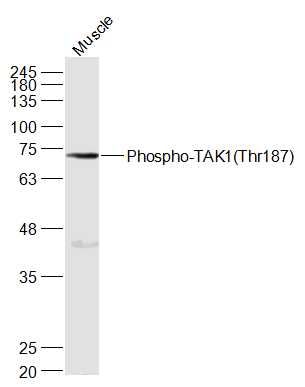
Sample:
Muscle (Mouse) Lysate at 40 ug
Primary: Anti-Phospho-TAK1(Thr187) (SL3438R) at 1/1000 dilution
Secondary: IRDye800CW Goat Anti-Rabbit IgG at 1/20000 dilution
Predicted band size: 67 kD
Observed band size: 70 kD
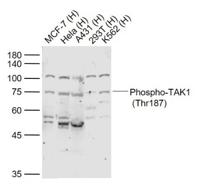
Sample:
Lane 1: MCF-7 (Human) Cell Lysate at 30 ug
Lane 2: Hela (Human) Cell Lysate at 30 ug
Lane 3: A431 (Human) Cell Lysate at 30 ug
Lane 4: 293T (Human) Cell Lysate at 30 ug
Lane 5: K562 (Human) Cell Lysate at 30 ug
Primary:
Anti-Phospho-TAK1 (Thr187) (SL3438R) at 1/1000 dilution
Secondary: IRDye800CW Goat Anti-Rabbit IgG at 1/20000 dilution
Predicted band size: 78 kD
Observed band size: 75 kD
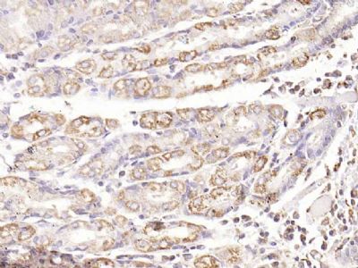
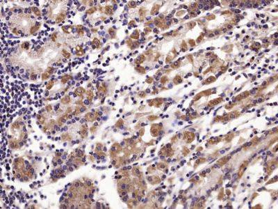
Paraformaldehyde-fixed, paraffin embedded (human gastric carcinoma); Antigen retrieval by boiling in sodium citrate buffer (pH6.0) for 15min; Block endogenous peroxidase by 3% hydrogen peroxide for 20 minutes; Blocking buffer (normal goat serum) at 37°C for 30min; Antibody incubation with (Phospho-TAK1 (Thr187)) Polyclonal Antibody, Unconjugated (SL3438R) at 1:200 overnight at 4°C, followed by operating according to SP Kit(Rabbit) (sp-0023) instructionsand DAB staining.
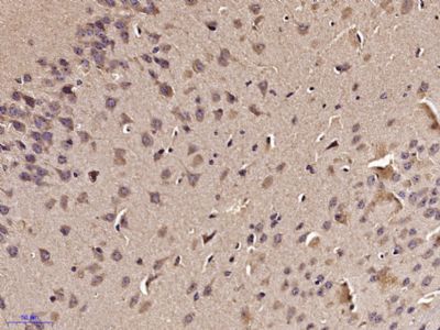
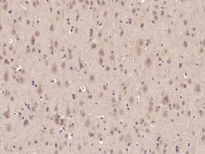
Paraformaldehyde-fixed, paraffin embedded (Mouse brain); Antigen retrieval by microwave in sodium citrate buffer (pH6.0) ; Block endogenous peroxidase by 3% hydrogen peroxide for 30 minutes; Blocking buffer (3% BSA) at RT for 30min; Antibody incubation with (Phospho-TAK1(Thr187)) Polyclonal Antibody, Unconjugated (SL3438R) at 1:400 overnight at 4℃, followed by conjugation to the secondary antibody (labeled with HRP)and DAB staining.
Paraformaldehyde-fixed, paraffin embedded (Human stomach); Antigen retrieval by microwave in sodium citrate buffer (pH6.0) ; Block endogenous peroxidase by 3% hydrogen peroxide for 30 minutes; Blocking buffer (3% BSA) at RT for 30min; Antibody incubation with (Phospho-TAK1(Thr187)) Polyclonal Antibody, Unconjugated (SL3438R) at 1:400 overnight at 4℃, followed by conjugation to the secondary antibody (labeled with HRP)and DAB staining.
Paraformaldehyde-fixed, paraffin embedded (Rat brain); Antigen retrieval by microwave in sodium citrate buffer (pH6.0) ; Block endogenous peroxidase by 3% hydrogen peroxide for 30 minutes; Blocking buffer (3% BSA) at RT for 30min; Antibody incubation with (Phospho-TAK1(Thr187)) Polyclonal Antibody, Unconjugated (SL3438R) at 1:400 overnight at 4℃, followed by conjugation to the secondary antibody (labeled with HRP)and DAB staining.
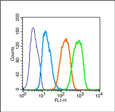
Blank control (Black line): Raji (Black).
Primary Antibody (green line): Rabbit Anti-Phospho-TAK1(Thr187) antibody (SL3438R)
Dilution: 1μg /10^6 cells;
Isotype Control Antibody (orange line): Rabbit IgG .
Secondary Antibody (white blue line): Goat anti-rabbit IgG-PE
Dilution: 1μg /test.
Protocol
The cells were fixed with 70% ice-cold methanol overnight at 4℃ and then permeabilized with 0.1% PBS-Tween for 20 min at room temperature (The cells were fixed with 2% paraformaldehyde (10 min) , then permeabilized with 90% ice-cold methanol for 20 min on ice.). Cells stained with Primary Antibody for 30 min at room temperature. The cells were then incubated in 1 X PBS/2%BSA/10% goat serum to block non-specific protein-protein interactions followed by the antibody for 15 min at room temperature. The secondary antibody used for 40 min at room temperature. Acquisition of 20,000 events was performed.
|