Sample:
HepG2(Human) Cell Lysate at 30 ug
Primary: Anti-Apolipoprotein E3 (SL5039R) at 1/2000 dilution
Secondary: IRDye800CW Goat Anti-Rabbit IgG at 1/20000 dilution
Predicted band size: 34 kD
Observed band size: 54 kD
Sample:
Plasma (Mouse) Lysate at 40 ug
Primary: Anti-Apolipoprotein E3 (SL5039R) at 1/1000 dilution
Secondary: IRDye800CW Goat Anti-Rabbit IgG at 1/20000 dilution
Predicted band size: 34 kD
Observed band size: 34 kD
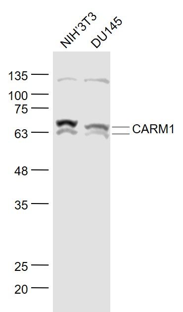
Sample:
Lane 1: Mouse Cerebellum Lysates
Lane 2: Mouse Brain Lysates
Primary: Anti-Apolipoprotein E3 (SL5039R) at 1/1000 dilution
Secondary: IRDye800CW Goat Anti-Rabbit IgG at 1/20000 dilution
Predicted band size: 34kDa
Observed band size: 35kDa
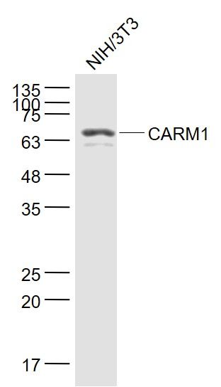
Sample:
Cerebrum (Mouse) Lysate at 40 ug
Cerebrum (Rat) Lysate at 40 ug
Primary: Anti-Apolipoprotein E3 (SL5039R) at 1/2000 dilution
Secondary: IRDye800CW Goat Anti-Rabbit IgG at 1/20000 dilution
Predicted band size: 34 kD
Observed band size: 49 kD
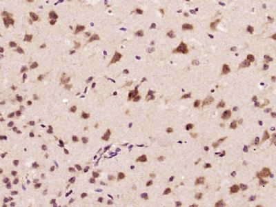
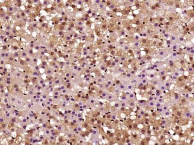
Tissue/cell: rat brain tissue; 4% Paraformaldehyde-fixed and paraffin-embedded;
Antigen retrieval: citrate buffer ( 0.01M, pH 6.0 ), Boiling bathing for 15min; Block endogenous peroxidase by 3% Hydrogen peroxide for 30min; Blocking buffer (normal goat serum,SLC0005) at 37℃ for 20 min;
Incubation: Anti-APOE3 Polyclonal Antibody, Unconjugated(SL5039R) 1:200, overnight at 4°C, followed by conjugation to the secondary antibody(SP-0023) and DAB(SLC0010) staining
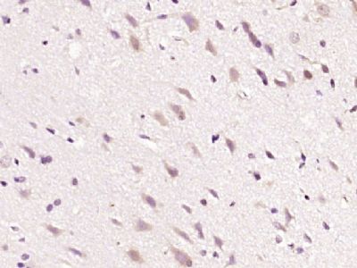
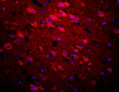
Tissue/cell: rat brain tissue;4% Paraformaldehyde-fixed and paraffin-embedded;
Antigen retrieval: citrate buffer ( 0.01M, pH 6.0 ), Boiling bathing for 15min; Blocking buffer (normal goat serum,SLC0005) at 37℃ for 20 min;
Incubation: Anti-APOE3 Polyclonal Antibody, Unconjugated(SL5039R) 1:200, overnight at 4°C; The secondary antibody was Goat Anti-Rabbit IgG, Cy3 conjugated(SL0295G-Cy3)used at 1:200 dilution for 40 minutes at 37°C. DAPI(5ug/ml,blue,SLC0033) was used to stain the cell nuclei
|