The immunosuppressant cyclosporin A (CsA) forms a trimolecular complex with cyclophilin and calcineurins to inhibit calcineurin phosphatase activity (1). Cyclophilins are conserved, ubiquitous and abundant cytosolic peptidyl-prolyl cis-trans isomerases that accelerate the isomerization of XaaPro peptide bonds and the refolding of proteins (2,3). Human cyclophilin A (CyPA), an intracellular protein of 165 amino acids, is the target of the CsA and is encoded by a single unique gene conserved from yeast to humans (4,5). CyPA is known for its involvement in T cell differentiation and proliferation and is highly expressed in brain (6). CyPA is incorporated into the virion of the type 1 human immunodeficiency virus (HISLV1) via a direct interaction with the capsid domain of the viral Gag polyprotein and is crucial for efficient viral replication (7,8). Cyclophilin B (CyPB) is a member of the cyclophilin family with specific N- and SLCterminal extensions. Unlike CyPA, CyPB has a signal sequence leading to its translocation in the endoplasmic reticulum. CyPB is secreted in biological fluids such as blood or milk and binds to a specific receptor present on the human lymphoblastic cell line Jurkat and on human peripheral blood lymphocytes (9,10).
Function:
PPIases accelerate the folding of proteins. It catalyzes the cis-trans isomerization of proline imidic peptide bonds in oligopeptides.
Subcellular Location:
Cytoplasm.
Similarity:
Belongs to the cyclophilin-type PPIase family. PPIase A subfamily.
Contains 1 PPIase cyclophilin-type domain.
SWISS:
P62937
Gene ID:
5478
Database links:
Entrez Gene: 5478 Human
Entrez Gene: 268373 Mouse
Entrez Gene: 10072977 Rat
Entrez Gene: 100362969 Rat
Entrez Gene: 25518 Rat
Omim: 123168 Human
SwissProt: P62937 Human
SwissProt: P17742 Mouse
SwissProt: P10111 Rat
Unigene: 356331 Human
Unigene: 598115 Human
Unigene: 5246 Mouse
Unigene: 118772 Rat
Unigene: 1463 Rat
| Picture |
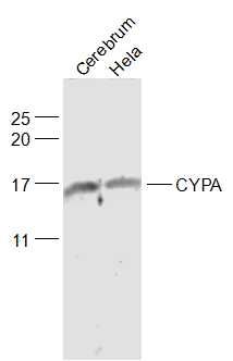
Sample:
Cerebrum (Rat) Lysate at 40 ug
Hela(Human) Cell Lysate at 30 ug
Primary: Anti-CYPA (SL5912R) at 1/1000 dilution
Secondary: IRDye800CW Goat Anti-Rabbit IgG at 1/20000 dilution
Predicted band size: 18 kD
Observed band size: 17/18 kD
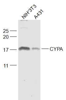
Sample:
NIH/3T3(Mouse) Cell Lysate at 30 ug
A431(Human) Cell Lysate at 30 ug
Primary: Anti-CYPA (SL5912R) at 1/1000 dilution
Secondary: IRDye800CW Goat Anti-Rabbit IgG at 1/20000 dilution
Predicted band size: 18 kD
Observed band size: 18 kD
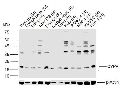
Sample:
Lane 1: Mouse Thymus tissue lysates
Lane 2: Mouse Lymph node tissue lysates
Lane 3: Mouse Lung tissue lysates
Lane 4: Mouse NIH/3T3 cell lysates
Lane 5: Rat Thymus tissue lysates
Lane 6: Rat Lymph node tissue lysates
Lane 7: Rat Lung tissue lysates
Lane 8: Human Hela cell lysates
Lane 9: Human PANSLC1 cell lysates
Lane 10: Human Molt-4 cell lysates
Lane 11: Human HUVEC cell lysates
Lane 12: Human THP-1 cell lysates
Primary: Anti- CYPA (SL5912R) at 1/1000 dilution
Secondary: IRDye800CW Goat Anti-Rabbit IgG at 1/20000 dilution
Predicted band size: 18 kDa
Observed band size: 18 kDa
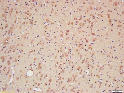
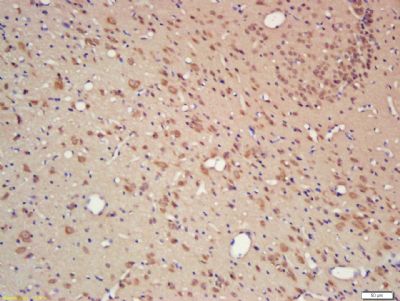
Tissue/cell: rat brain tissue; 4% Paraformaldehyde-fixed and paraffin-embedded;
Antigen retrieval: citrate buffer ( 0.01M, pH 6.0 ), Boiling bathing for 15min; Block endogenous peroxidase by 3% Hydrogen peroxide for 30min; Blocking buffer (normal goat serum,SLC0005) at 37℃ for 20 min;
Incubation: Anti-CYPA Polyclonal Antibody, Unconjugated(SL5912R) 1:200, overnight at 4°C, followed by conjugation to the secondary antibody(SP-0023) and DAB(SLC0010) staining
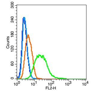
Blank control(blue): Hela(fixed with 2% paraformaldehyde (10 min) , then permeabilized with 90% ice-cold methanol for 30 min on ice).
Primary Antibody:Rabbit Anti- CYPA antibody(SL5912R), Dilution: 1μg in 100 μL 1X PBS containing 0.5% BSA;
Isotype Control Antibody: Rabbit IgG(orange) ,used under the same conditions );
Secondary Antibody: Goat anti-rabbit IgG-PE(white blue), Dilution: 1:200 in 1 X PBS containing 0.5% BSA.
|
|
|