Home > Product > Antibody > Rabbit Anti-CAMKK2 antibody
Calcium/calmodulin dependent protein kinase beta; Calcium/calmodulin dependent protein kinase kinase 2; Calcium/calmodulin dependent protein kinase kinase beta; CaM kinase kinase beta; CaM KK beta; CAMKK 2; CAMKK; CaMKK beta; CAMKK beta protein; CAMKKB.
Cat:
SL6253R
Species Reactivity:
Human,Mouse,Rat,Sheep,(predicted: Pig,Cow,Horse,)
Immunogen:
KLH conjugated synthetic peptide derived from human CAMKK2:501-588/588
Format:
Liquid
Storage instructions:
Shipped at 4℃. Store at -20 °C for one year. Avoid repeated freeze/thaw cycles.
Concentration:
1mg/ml
Clonality:
Polyclonal
Isotype:
IgG
Applications:
WB=1:500-2000IHC-P=1:100-500IHC-F=1:100-500Flow-Cyt=1ug/Test(Paraffin sections need to do antigen repair)not yet tested in other applications.optimal dilutions/concentrations should be determined by the end user.
Host:
Rabbit
Product Overview:
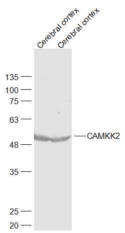
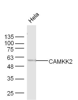
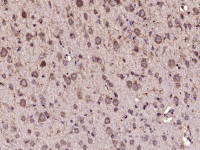
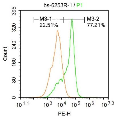
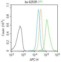
Sample: Cerebral cortex (Mouse) Lysate at 40 ugCerebral cortex (Rat) Lysate at 40 ugPrimary: Anti-CAMKK2 (SL6253R) at 1/1000 dilutionSecondary: IRDye800CW Goat Anti-Rabbit IgG at 1/20000 dilutionPredicted band size: 55 kDObserved band size: 55 kDSample: Hela Cell (Human) Lysate at 40 ugPrimary: Anti-CAMKK2 (SL6253R) at 1/300 dilutionSecondary: IRDye800CW Goat Anti-Rabbit IgG at 1/20000 dilutionPredicted band size: 55 kDObserved band size: 55 kD Paraformaldehyde-fixed, paraffin embedded (Rat brain); Antigen retrieval by microwave in sodium citrate buffer (pH6.0) ; Block endogenous peroxidase by 3% hydrogen peroxide for 30 minutes; Blocking buffer (3% BSA) at RT for 30min; Antibody incubation with (CAMKK2) Polyclonal Antibody, Unconjugated (SL6253R) at 1:400 overnight at 4°C, followed by conjugation to the secondary antibody (labeled with HRP)and DAB staining.Molt-4 cells were fixed with 4% PFA for 10min at room temperature,permeabilized with 0.1% PBST for 20 min at room temperature, and incubated in 5% BSA blocking buffer for 30 min at room temperature. Cells were then stained with CAMKK2 Antibody(SL6253R)at 1:100 dilution in blocking buffer and incubated for 30 min at room temperature, washed twice with 2%BSA in PBS, followed by secondary antibody incubation for 40 min at room temperature. Acquisitions of 20,000 events were performed. Cells stained with primary antibody (green), and isotype control (orange).Blank control (Black line): Molt4 (Black). Primary Antibody (green line):Rabbit Anti-CAMKK2 antibody (SL6253R)Dilution: 1μg /10^6 cells; Isotype Control Antibody (orange line): Rabbit IgG . Secondary Antibody (white blue line): Goat anti-rabbit IgG-AF647Dilution: 1μg /test. ProtocolThe cells were fixed with 4% PFA (10min at room temperature)and then permeabilized with PBST for 20 min at room temperature. The cells were then incubated in 5%BSA to block non-specific protein-protein interactions for 30 min at room temperature .Cells stained with Primary Antibody for 30 min at room temperature. The secondary antibody used for 40 min at room temperature. Acquisition of 20,000 events was performed.
More
Unit:
Price: $
Product PDFs
Datasheet:


The product of this gene belongs to the Serine/Threonine protein kinase family, and to the Ca(2+)/calmodulin-dependent protein kinase subfamily. This protein plays a role in the calcium/calmodulin-dependent (CaM) kinase cascade by phosphorylating the downstream kinases CaMK1 and CaMK4. Seven transcript variants encoding six distinct isoforms have been identified for this gene. Additional splice variants have been described but their full-length nature has not been determined. The identified isoforms exhibit a distinct ability to undergo autophosphorylation and to phosphorylate the downstream kinases. [provided by RefSeq, Jul 2008].

Function:
Calcium/calmodulin-dependent protein kinase belonging to a proposed calcium-triggered signaling cascade involved in a number of cellular processes. Isoform 1, isoform 2 and isoform 3 phosphorylate CAMK1 and CAMK4. Isoform 3 phosphorylates CAMK1D. Isoform 4, isoform 5 and isoform 6 lacking part of the calmodulin-binding domain are inactive. Efficiently phosphorylates 5'-AMP-activated protein kinase (AMPK) trimer, including that consisting of PRKAA1, PRKAB1 and PRKAG1. This phosphorylation is stimulated in response to Ca(2+) signals (By similarity). Seems to be involved in hippocampal activation of CREB1 (By similarity). May play a role in neurite growth. Isoform 3 may promote neurite elongation, while isoform 1 may promoter neurite branching.

Subunit:
Interacts with calmodulin.

Subcellular Location:
Nucleus. Cytoplasm. Cell projection. Note=Predominantly nuclear in unstimulated cells. Found in the cytoplasm and neurites after forskolin induction.

Tissue Specificity:
Ubiquitously expressed with higher levels in the brain. Intermediate levels are detected in spleen, prostate, thyroid and leukocytes. The lowest level is in lung.

Post-translational modifications:
Autophosphorylated and phosphorylated by PKA. Each isoform may show a different pattern of phosphorylation.

Similarity:
Belongs to the protein kinase superfamily. Ser/Thr protein kinase family.
Contains 1 protein kinase domain.

SWISS:
Q96RR4

Gene ID:
10645

Database links:

Entrez Gene: 10645 Human

Entrez Gene: 83506 Rat

Omim: 63002 Human

SwissProt: Q96RR4 Human

SwissProt: O88831 Rat

Unigene: 297343 Human

Unigene: 88589 Rat


Picture

Sample:
Cerebral cortex (Mouse) Lysate at 40 ug
Cerebral cortex (Rat) Lysate at 40 ug
Primary: Anti-CAMKK2 (SL6253R) at 1/1000 dilution
Secondary: IRDye800CW Goat Anti-Rabbit IgG at 1/20000 dilution
Predicted band size: 55 kD
Observed band size: 55 kD
Sample: Hela Cell (Human) Lysate at 40 ug
Primary: Anti-CAMKK2 (SL6253R) at 1/300 dilution
Secondary: IRDye800CW Goat Anti-Rabbit IgG at 1/20000 dilution
Predicted band size: 55 kD
Observed band size: 55 kD
Paraformaldehyde-fixed, paraffin embedded (Rat brain); Antigen retrieval by microwave in sodium citrate buffer (pH6.0) ; Block endogenous peroxidase by 3% hydrogen peroxide for 30 minutes; Blocking buffer (3% BSA) at RT for 30min; Antibody incubation with (CAMKK2) Polyclonal Antibody, Unconjugated (SL6253R) at 1:400 overnight at 4°C, followed by conjugation to the secondary antibody (labeled with HRP)and DAB staining.
Molt-4 cells were fixed with 4% PFA for 10min at room temperature,permeabilized with 0.1% PBST for 20 min at room temperature, and incubated in 5% BSA blocking buffer for 30 min at room temperature. Cells were then stained with CAMKK2 Antibody(SL6253R)at 1:100 dilution in blocking buffer and incubated for 30 min at room temperature, washed twice with 2%BSA in PBS, followed by secondary antibody incubation for 40 min at room temperature. Acquisitions of 20,000 events were performed. Cells stained with primary antibody (green), and isotype control (orange).
Blank control (Black line): Molt4 (Black).
Primary Antibody (green line):Rabbit Anti-CAMKK2 antibody (SL6253R)
Dilution: 1μg /10^6 cells;
Isotype Control Antibody (orange line): Rabbit IgG .
Secondary Antibody (white blue line): Goat anti-rabbit IgG-AF647
Dilution: 1μg /test.
Protocol
The cells were fixed with 4% PFA (10min at room temperature)and then permeabilized with PBST for 20 min at room temperature. The cells were then incubated in 5%BSA to block non-specific protein-protein interactions for 30 min at room temperature .Cells stained with Primary Antibody for 30 min at room temperature. The secondary antibody used for 40 min at room temperature. Acquisition of 20,000 events was performed.
Product Feedback Wall
Message :
Your Email :
Copyright © 2007-2018 Sunlong Medical All Rights Reserved.