Home > Product > Antibody > Rabbit Anti-LGI1 antibody
Epitempin 1; EPITEMPIN; Epitempin-1; EPT; ETL1; IB1099; Leucine rich glioma-inactivated protein 1; Leucine-rich glioma-inactivated protein 1; LGI1; LGI1_HUMAN; EPITEMPIN.
Cat:
SL6719R
Species Reactivity:
Mouse,Rat,(predicted: Human,Dog,Pig,Cow,Horse,Rabbit,)
Immunogen:
KLH conjugated synthetic peptide derived from human LGI1/ETL1:451-557/557
Format:
Liquid
Storage instructions:
Shipped at 4℃. Store at -20 °C for one year. Avoid repeated freeze/thaw cycles.
Concentration:
1mg/ml
Clonality:
Polyclonal
Isotype:
IgG
Applications:
WB=1:500-2000ELISA=1:5000-10000IHC-P=1:100-500IHC-F=1:100-500IF=1:100-500(Paraffin sections need to do antigen repair)not yet tested in other applications.optimal dilutions/concentrations should be determined by the end user.
Host:
Rabbit
Product Overview:
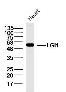
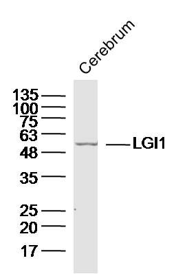
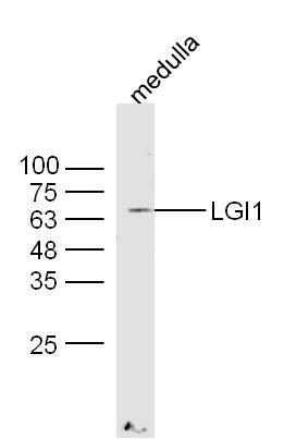
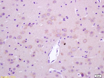
Sample:Heart (Mouse) Lysate at 40 ugPrimary: Anti-LGI1(SL6719R)at 1/300 dilutionSecondary: IRDye800CW Goat Anti-Rabbit IgG at 1/20000 dilutionPredicted band size: 64kDObserved band size: 60kDSample:Cerebrum (Mouse) Lysate at 40 ugPrimary: Anti-LGI1(SL6719R)at 1/300 dilutionSecondary: IRDye800CW Goat Anti-Rabbit IgG at 1/20000 dilutionPredicted band size: 64kDObserved band size: 60kDSample: Medulla (Mouse) Lysate at 40 ugPrimary: Anti-LGI1 (SL6719R) at 1/300 dilutionSecondary: IRDye800CW Goat Anti-Rabbit IgG at 1/20000 dilutionPredicted band size: 64 kDObserved band size: 64 kDParaformaldehyde-fixed, paraffin embedded (rat brain tissue); Antigen retrieval by boiling in sodium citrate buffer (pH6.0) for 15min; Block endogenous peroxidase by 3% hydrogen peroxide for 20 minutes; Blocking buffer (normal goat serum) at 37°C for 30min; Antibody incubation with (LGI1) Polyclonal Antibody, Unconjugated (SL6719R) at 1:400 overnight at 4°C, followed by operating according to SP Kit(Rabbit) (sp-0023) instructionsand DAB staining.Tissue/cell: rat brain tissue; 4% Paraformaldehyde-fixed and paraffin-embedded; Antigen retrieval: citrate buffer ( 0.01M, pH 6.0 ), Boiling bathing for 15min; Block endogenous peroxidase by 3% Hydrogen peroxide for 30min; Blocking buffer (normal goat serum,SLC0005) at 37℃ for 20 min; Incubation: Anti-LGI1/ETL1 Polyclonal Antibody, Unconjugated(SL6719R) 1:200, overnight at 4°C, followed by conjugation to the secondary antibody(SP-0023) and DAB(SLC0010) staining
More
Unit:
Price: $
Product PDFs
Datasheet:


Regulates voltage-gated potassium channels assembled from KCNA1, KCNA4 and KCNAB1. It slows down channel inactivation by precluding channel closure mediated by the KCNAB1 subunit. Ligand for ADAM22 that positively regulates synaptic transmission mediated by AMPA-type glutamate receptors (By similarity). Plays a role in suppressing the production of MMP1/3 through the phosphatidylinositol 3-kinase/ERK pathway. May play a role in the control of neuroblastoma cell survival.
Tissue specificity;Predominantly expressed in neural tissues, especially in brain. Expression is reduced in low-grade brain tumors and significantly reduced or absent in malignant gliomas. Isoform 1 is absent in the cerebellum and is detectable in the occipital cortex and hippocampus; higher amounts are observed in the parietal and frontal cortices, putamen, and, particularly, in the temporal neocortex, where it is 3.5 times more abundant than in the hippocampus (at protein level). Isoform 3 shows the highest expression in the occipital cortex and the lowest in the hippocampus (at protein level).

Function:
Regulates voltage-gated potassium channels assembled from KCNA1, KCNA4 and KCNAB1. It slows down channel inactivation by precluding channel closure mediated by the KCNAB1 subunit. Ligand for ADAM22 that positively regulates synaptic transmission mediated by AMPA-type glutamate receptors (By similarity). Plays a role in suppressing the production of MMP1/3 through the phosphatidylinositol 3-kinase/ERK pathway. May play a role in the control of neuroblastoma cell survival.

Subunit:
Oligomer (By similarity). Interacts with KCNA1 within a complex containing KCNA1, KCNA4 and KCNAB1 (By similarity). Part of a complex containing ADAM22, DLG4/PSD95 and CACNG2 (stargazin) (By similarity). Can bind to ADAM11 and ADAM23 (By similarity).

Subcellular Location:
Secreted. Cell junction, synapse (By similarity). Note=Isoform 1 but not isoform 2 is secreted. Isoform 1 is enriched in the Golgi apparatus while isoform 2 accumulates in the endoplasmic reticulum.

Tissue Specificity:
Predominantly expressed in neural tissues, especially in brain. Expression is reduced in low-grade brain tumors and significantly reduced or absent in malignant gliomas. Isoform 1 is absent in the cerebellum and is detectable in the occipital cortex and hippocampus; higher amounts are observed in the parietal and frontal cortices, putamen, and, particularly, in the temporal neocortex, where it is 3.5 times more abundant than in the hippocampus (at protein level). Isoform 3 shows the highest expression in the occipital cortex and the lowest in the hippocampus (at protein level).

Post-translational modifications:
Glycosylated.

DISEASE:
Defects in LGI1 are the cause of lateral temporal lobe epilepsy autosomal dominant (ADLTE) [MIM:600512]; also known as autosomal dominant partial epilepsy with auditory features (ADPEAF). ADLTE is a form of epilepsy characterized by partial seizures, usually preceded by auditory signs.

Similarity:
Contains 7 EAR repeats.
Contains 3 LRR (leucine-rich) repeats.
Contains 1 LRRCT domain.
Contains 1 LRRNT domain

SWISS:
O95970

Gene ID:
9211

Database links:

Entrez Gene: 9211 Human

Entrez Gene: 56839 Mouse

Entrez Gene: 252892 Rat

Omim: 604619 Human

SwissProt: O95970 Human

SwissProt: Q9JIA1 Mouse

SwissProt: Q8K4Y5 Rat

Unigene: 533670 Human

Unigene: 298251 Mouse

Unigene: 83615 Rat


Picture

Sample:Heart (Mouse) Lysate at 40 ug
Primary: Anti-LGI1(SL6719R)at 1/300 dilution
Secondary: IRDye800CW Goat Anti-Rabbit IgG at 1/20000 dilution
Predicted band size: 64kD
Observed band size: 60kD
Sample:Cerebrum (Mouse) Lysate at 40 ug
Primary: Anti-LGI1(SL6719R)at 1/300 dilution
Secondary: IRDye800CW Goat Anti-Rabbit IgG at 1/20000 dilution
Predicted band size: 64kD
Observed band size: 60kD
Sample: Medulla (Mouse) Lysate at 40 ug
Primary: Anti-LGI1 (SL6719R) at 1/300 dilution
Secondary: IRDye800CW Goat Anti-Rabbit IgG at 1/20000 dilution
Predicted band size: 64 kD
Observed band size: 64 kD
Paraformaldehyde-fixed, paraffin embedded (rat brain tissue); Antigen retrieval by boiling in sodium citrate buffer (pH6.0) for 15min; Block endogenous peroxidase by 3% hydrogen peroxide for 20 minutes; Blocking buffer (normal goat serum) at 37°C for 30min; Antibody incubation with (LGI1) Polyclonal Antibody, Unconjugated (SL6719R) at 1:400 overnight at 4°C, followed by operating according to SP Kit(Rabbit) (sp-0023) instructionsand DAB staining.
Tissue/cell: rat brain tissue; 4% Paraformaldehyde-fixed and paraffin-embedded;
Antigen retrieval: citrate buffer ( 0.01M, pH 6.0 ), Boiling bathing for 15min; Block endogenous peroxidase by 3% Hydrogen peroxide for 30min; Blocking buffer (normal goat serum,SLC0005) at 37℃ for 20 min;
Incubation: Anti-LGI1/ETL1 Polyclonal Antibody, Unconjugated(SL6719R) 1:200, overnight at 4°C, followed by conjugation to the secondary antibody(SP-0023) and DAB(SLC0010) staining
Product Feedback Wall
Message :
Your Email :
Copyright © 2007-2018 Sunlong Medical All Rights Reserved.