The protein encoded by this gene is a transcriptional coactivator that regulates the genes involved in energy metabolism. This protein interacts with PPARgamma, which permits the interaction of this protein with multiple transcription factors. This protein can interact with, and regulate the activities of, cAMP response element binding protein (CREB) and nuclear respiratory factors (NRFs). It provides a direct link between external physiological stimuli and the regulation of mitochondrial biogenesis, and is a major factor that regulates muscle fiber type determination. This protein may be also involved in controlling blood pressure, regulating cellular cholesterol homoeostasis, and the development of obesity. [provided by RefSeq].
Function:
Transcriptional coactivator for steroid receptors and nuclear receptors. Greatly increases the transcriptional activity of PPARG and thyroid hormone receptor on the uncoupling protein promoter. Can regulate key mitochondrial genes that contribute to the program of adaptive thermogenesis.
Subunit:
Binds MYBBP1A, which inhibits transcriptional activation by this protein. Interacts with PRDM16. Interacts with LRPPRC. Homooligomer. Interacts with LPIN1.
Subcellular Location:
Nucleus.
Tissue Specificity:
Heart, skeletal muscle, liver and kidney. Expressed at lower levels in brain and pancreas and at very low levels in the intestine and white adipose tissue. In skeletal muscle, levels were lower in obese than in lean subjects and fasting induced a 2-fold increase in levels in the skeletal muscle in obese subjects.
Post-translational modifications:
Phosphorylation by AMPK in skeletal muscle increases activation of its own promoter. Phosphorylated by CLK2.
Similarity:
Contains 1 RRM (RNA recognition motif) domain.
SWISS:
Q9UBK2
Gene ID:
10891
Database links:
Entrez Gene: 10891 Human
Entrez Gene: 133522 Human
Entrez Gene: 170826 Mouse
Entrez Gene: 19017 Mouse
Entrez Gene: 291567 Rat
Entrez Gene: 83516 Rat
Omim: 604517 Human
Omim: 608886 Human
SwissProt: Q86YN6 Human
SwissProt: Q9UBK2 Human
SwissProt: 415302 Mouse
SwissProt: O70343 Mouse
SwissProt: Q8VHJ7 Mouse
SwissProt: Q811R2 Rat
SwissProt: Q9QYK2 Rat
Unigene: 483816 Human
Unigene: 527078 Human
Unigene: 163382 Rat
| Picture |
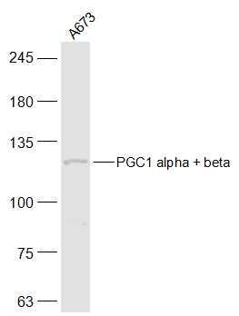
Sample:
A673(Human) Cell Lysate at 30 ug
Primary: Anti-PGC1 alpha + beta (SL7535R) at 1/500 dilution
Secondary: IRDye800CW Goat Anti-Rabbit IgG at 1/20000 dilution
Predicted band size: 88/113 kD
Observed band size: 113 kD
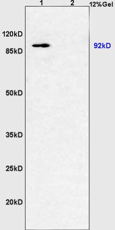
Sample:
Muscle (Pig) Lysate at 40 ug
Kidney (Mouse) Lysate at 40 ug
Primary: Anti-PGC1 alpha + beta (SL4064R) at 1/300 dilution
Secondary: HRP conjugated Goat-Anti-rabbit IgG (SL0295G-HRP) at 1/5000 dilution
Predicted band size: 88/113 kD
Observed band size: 92 kD
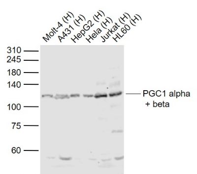
Sample:
Lane 1: Molt-4 (Human) Cell Lysate at 30 ug
Lane 2: A431 (Human) Cell Lysate at 30 ug
Lane 3: HepG2 (Human) Cell Lysate at 30 ug
Lane 4: Hela (Human) Cell Lysate at 30 ug
Lane 5: Jurkat (Human) Cell Lysate at 30 ug
Lane 6: HL60 (Human) Cell Lysate at 30 ug
Primary: Anti-PGC1 alpha + beta (SL7535R) at 1/1000 dilution
Secondary: IRDye800CW Goat Anti-Rabbit IgG at 1/20000 dilution
Predicted band size: 90/113 kD
Observed band size: 113 kD
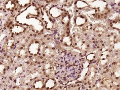
Paraformaldehyde-fixed, paraffin embedded (rat kidney tissue); Antigen retrieval by boiling in sodium citrate buffer (pH6.0) for 15min; Block endogenous peroxidase by 3% hydrogen peroxide for 20 minutes; Blocking buffer (normal goat serum) at 37°C for 30min; Antibody incubation with (PGC1 alpha + beta) Polyclonal Antibody, Unconjugated (SL7535R) at 1:400 overnight at 4°C, followed by operating according to SP Kit(Rabbit) (sp-0023) instructionsand DAB staining.
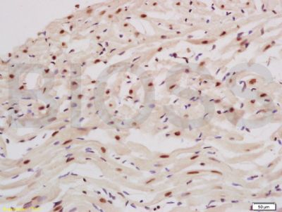
Tissue/cell: rat heart tissue; 4% Paraformaldehyde-fixed and paraffin-embedded;
Antigen retrieval: citrate buffer ( 0.01M, pH 6.0 ), Boiling bathing for 15min; Block endogenous peroxidase by 3% Hydrogen peroxide for 30min; Blocking buffer (normal goat serum,SLC0005) at 37℃ for 20 min;
Incubation: Anti-PGC1 alpha+beta Polyclonal Antibody, Unconjugated(SL7535R) 1:200, overnight at 4°C, followed by conjugation to the secondary antibody(SP-0023) and DAB(SLC0010) staining
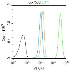
Blank control (Black line): Molt4 (Black).
Primary Antibody (green line):Rabbit Anti-PGC1 alpha+beta antibody (SL7535R)
Dilution:1μg /10^6 cells;
Isotype Control Antibody (orange line): Rabbit IgG .
Secondary Antibody (white blue line): Goat anti-rabbit IgG-AF647
Dilution: 1μg /test.
Protocol
The cells were fixed with 4% PFA (10min at room temperature)and then permeabilized with 90% ice-cold methanol for 20 min at room temperature. The cells were then incubated in 5%BSA to block non-specific protein-protein interactions for 30 min at room temperature .Cells stained with Primary Antibody for 30 min at room temperature. The secondary antibody used for 40 min at room temperature. Acquisition of 20,000 events was performed.
|
|
|