[IF=3.895] Yang W et al. The Effect of Low and High Dose Deoxynivalenol on Intestinal Morphology, Distribution, and Expression of Inflammatory Cytokines of Weaning Rabbits. Toxins (Basel). 2019 Aug 13;11(8). pii: E473. WB&IHSLCP ; Rabbit.
[IF=2.882] Gul Fatma Yarim. et al. Nobiletin attenuates inflammation via modulating proinflammatory and antiinflammatory cytokine expressions in an autoimmune encephalomyelitis mouse model. Fitoterapia. 2022 Jan;156:105099 IHC ; Rat.
[IF=11.799] Chenyu Zhang. et al. The Nrf2-NLRP3-caspase-1 axis mediates the neuroprotective effects of Celastrol in Parkinson's disease. Redox Biol. 2021 Nov;47:102134 WB ; mosue.
[IF=3.412] Abdelaziz Adam Idriss Arbab. et al. Metformin Inhibits Lipoteichoic Acid–Induced Oxidative Stress and Inflammation Through AMPK/NRF2/NF-κB Signaling Pathway in Bovine Mammary Epithelial Cells. Front Vet Sci. 2021; 8: 66276 WB ; Bovine.
[IF=1.25] Mehmet Balbaba. et al. Anti-inflammatory effect of cortistatin in rat endotoxin-induced uveitis model. Indian J Ophthalmol. 2020 Sep; 68(9): 1920–1924 IHC ; Rat.
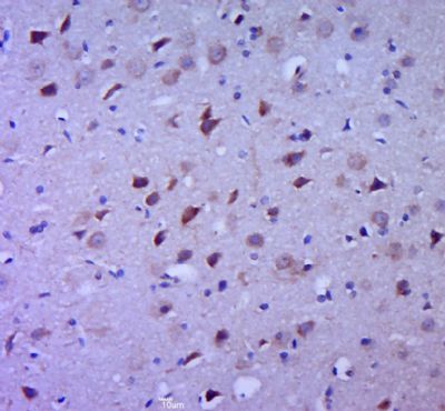
[IF=7.238] Chinthalapally V. Rao. et al. GSK3‐ARC/Arg3.1 and GSK3‐Wnt signaling axes trigger amyloid‐β accumulation and neuroinflammation in middle‐aged Shugoshin 1 mice. Aging Cell. 2020 Oct;19(10):e13221 IHC ; Mouse.
[IF=3.69] Dai-Chao Ma. et al. Salvianolic Acids for Injection alleviates cerebral ischemia/reperfusion injury by switching M1/M2 phenotypes and inhibiting NLRP3 inflammasome/pyroptosis axis in microglia in vivo and in vitro. J Ethnopharmacol. 2021 Apr;270:113776 WB ; Rat.
[IF=0] Yamada H et al. LATE-ONSET ALZHEIMER'S DISEASE ANIMAL MODEL AND USES THEREOF. US Patent20190387723 WB, IHSLCP&IHF ; Mouse.
[IF=4.13] Fukuda M et al. Disruption of P2X4 purinoceptor and suppression of the inflammation associated with cerebral aneurysm formation. J Neurosurg. 2019 Dec 20:1-13. WB ; Mouse.
[IF=3.17] Yu H et al. Echinocystic acid, a natural plant extract, alleviates cerebral ischemia/reperfusion injury via inhibiting the JNK signaling pathway. Eur J Pharmacol. 2019 Oct 15;861:172610. WB ; Mouse.
[IF=3.895] Sun D et al. Canonical Transient Receptor Potential Channel 3 Contributes to Febrile Seizure Inducing Neuronal Cell Death and Neuroinflammation.Cell. Mol. Neurobiol. 2018 Aug;38(6), 1215–1226. IF ; Rat.
[IF=2.55] Dinesh, Palani, and MahaboobKhan Rasool. "Berberine, an isoquinoline alkaloid suppresses TXNIP mediated NLRP3 inflammasome activation in MSU crystal stimulated RAW 264.7 macrophages through the upregulation of Nrf2 transcription factor and alleviates MSU crystal induced inflammation in rats." International Immunopharmacology 44 (2017): 26-37. WB ; Mouse.
[IF=3.921] Meiyun Cai. et al. Kaemperfol alleviates pyroptosis and microglia-mediated neuroinflammation in Parkinson's disease via inhibiting p38MAPK/NF-κB signaling pathway. Neurochem Int. 2021 Nov;:105221 WB ; Rat.
[IF=2.695] Jaclyn Iannucci. et al. Isoform-Specific Effects of Apolipoprotein E on Markers of Inflammation and Toxicity in Brain Glia and Neuronal Cells In Vitro. Curr Issues Mol Biol. 2021 Jun;43(1):215-225 WB ; Human.
[IF=4.858] Kun Hao. et al. Targeting BRD4 prevents acute gouty arthritis by regulating pyroptosis. Int J Biol Sci. 2020; 16(16): 3163–3173 WB ; Rat, Human.
[IF=4.966] Wei‐Cheng Chen. et al. Resistin enhances IL‐1β and TNF‐α expression in human osteoarthritis synovial fibroblasts by inhibiting miR‐149 expression via the MEK and ERK pathways. Faseb J. 2020 Oct;34(10):13671-13684 WB,IHC ; Human.
[IF=2.985] Mengmeng Yang. et al. Mesenchymal stem cell-conditioned medium improved mitochondrial function and alleviated inflammation and apoptosis in non-alcoholic fatty liver disease by regulating SIRT1. Biochem Bioph Res Co. 2021 Mar;546:74 WB ; Mouse, Human.
[IF=13.116] Zhang X et al. Oligodendroglial glycolytic stress triggers inflammasome activation and neuropathology in Alzheimer’s diseaseSci Adv.2020 Dec 4;6(49):eabb8136. IF ; Mouse.
[IF=4.691] Dai-Chao Maet al. Kv1.3 channel blockade alleviates cerebral ischemia/reperfusion injury by reshaping M1/M2 phenotypes and compromising the activation of NLRP3 inflammasome in microglia. Exp Neurol
. 2020 Oct;332:113399. WB ; rat.
[IF=6.306] Zhou P et al. Histamine-4 receptor antagonist JNJ7777120 inhibits pro-inflammatory microglia and prevents the progression of Parkinson-like pathology and behaviour in a rat model.(2018)Brain Behav. Immun. Nov 05. WB ; Rat.
[IF=1.41] Han JK et al. Anti-inflammatory effect of polydeoxyribonucleotide on zoledronic acid-pretreated and lipopolysaccharide-stimulated RAW 264.7 cells. (2018) Exp.Ther.Med. 16:400-405 WB ; human.
[IF=0] Evashwick-Rogler et al. Inhibiting tumor necrosis factor-alpha at time of induced intervertebral disc injury limits long-term pain and degeneration in a rat model. (2018) JOR.Spine. 1 IHC ; Rat.
[IF=2.33] Li, Kang, et al. "CD14 knockdown reduces lipopolysaccharide-induced cell viability and expression of inflammation-associated genes in gastric cancer cells in vitro and in nude mouse xenografts." Molecular Medicine Reports. WB ; Human.