Bardet-Biedl syndrome (BBS) is a pleiotropic genetic disorder characterized by obesity, photoreceptor degeneration, polydactyly, hypogenitalism, renal abnormalities, and developmental delay. BBS patients also have an increased risk of developing diabetes, hypertension, and congenital heart defects. BBS is a heterogeneous disorder mapping to eight genetic loci and encoding eight proteins, BBS1-BBS8. Five BBS proteins encode basal body or cilia proteins, suggesting that BBS is a ciliary dysfunction disorder. BBS2 contains two overlapping genes: BBS2L1 and BBS2L2. BBSL1 was re-named BBS7, whereas BBS2L2 independently funcitons as BBS1. BBS7 contains 672 amino acids and is expressed at low to moderate levels in most human tissues.
Function:
The BBSome complex is required for ciliogenesis but is dispensable for centriolar satellite function. This ciliogenic function is mediated in part by the Rab8 GDP/GTP exchange factor, which localizes to the basal body and contacts the BBSome. Rab8(GTP) enters the primary cilium and promotes extension of the ciliary membrane. Firstly the BBSome associates with the ciliary membrane and binds to RAB3IP/Rabin8, the guanosyl exchange factor (GEF) for Rab8 and then the Rab8-GTP localizes to the cilium and promotes docking and fusion of carrier vesicles to the base of the ciliary membrane.
Subunit:
Part of BBSome complex, that contains BBS1, BBS2, BBS4, BBS5, BBS7, BBS8, BBS9 and BBIP10. The BBSome complex binds to PCM1 and tubulin. Interacts with PCM1. Interacts with CCDC28B.
Subcellular Location:
Cytoplasm, cytoskeleton, centrosome. Cell projection, cilium membrane. Cytoplasm. Note=Localizes to nonmembranous centriolar satellites in the cytoplasm.
Tissue Specificity:
Widely expressed.
DISEASE:
Defects in TTC8 are the cause of retinitis pigmentosa type 51 (RP51) [MIM:613464]. It is a retinal dystrophy belonging to the group of pigmentary retinopathies. Retinitis pigmentosa is characterized by retinal pigment deposits visible on fundus examination and primary loss of rod photoreceptor cells followed by secondary loss of cone photoreceptors. Patients typically have night vision blindness and loss of midperipheral visual field. As their condition progresses, they lose their far peripheral visual field and eventually central vision as well.
Defects in TTC8 are the cause of Bardet-Biedl syndrome type 8 (BBS8) [MIM:209900]. Bardet-Biedl syndrome (BBS) is a genetically heterogeneous, autosomal recessive disorder characterized by usually severe pigmentary retinopathy, early onset obesity, polydactyly, hypogenitalism, renal malformation and mental retardation. Secondary features include diabetes mellitus, hypertension and congenital heart disease. A relatively high incidence of BBS is found in the mixed Arab populations of Kuwait and in Bedouin tribes throughout the Middle East, most likely due to the high rate of consaguinity in these populations and a founder effect.
Similarity:
Contains 8 TPR repeats.
SWISS:
Q8TAM2
Gene ID:
123016
Database links:
Entrez Gene: 123016 Human
Entrez Gene: 76260 Mouse
GenBank: NP_938051.1 Human
Omim: 608132 Human
SwissProt: Q8TAM2 Human
SwissProt: Q8VD72 Mouse
Unigene: 303055 Human
Unigene: 282660 Mouse
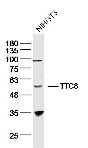
| Picture |
Sample: NIH/3T3 (human)cell Lysate at 40 ug
Primary: Anti- TTC8 (SL11510R)at 1/300 dilution
Secondary: IRDye800CW Goat Anti-Rabbit IgG at 1/20000 dilution
Predicted band size: 61kD
Observed band size: 58 kD
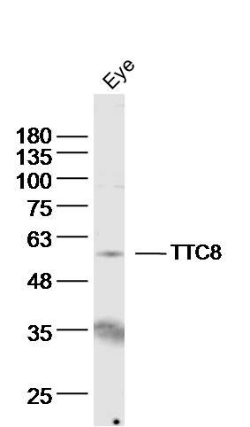
Sample: eye (mouse) Lysate at 40 ug
Primary: Anti- TTC8 (SL11510R)at 1/300 dilution
Secondary: IRDye800CW Goat Anti-Rabbit IgG at 1/20000 dilution
Predicted band size: 61kD
Observed band size: 58 kD
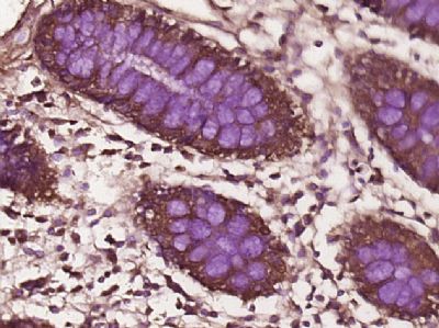
Paraformaldehyde-fixed, paraffin embedded (Human colon cancer); Antigen retrieval by boiling in sodium citrate buffer (pH6.0) for 15min; Block endogenous peroxidase by 3% hydrogen peroxide for 20 minutes; Blocking buffer (normal goat serum) at 37°C for 30min; Antibody incubation with (TTC8) Polyclonal Antibody, Unconjugated (SL11510R) at 1:400 overnight at 4°C, followed by operating according to SP Kit(Rabbit) (sp-0023) instructionsand DAB staining.
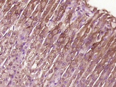
Paraformaldehyde-fixed, paraffin embedded (Mouse stomach); Antigen retrieval by boiling in sodium citrate buffer (pH6.0) for 15min; Block endogenous peroxidase by 3% hydrogen peroxide for 20 minutes; Blocking buffer (normal goat serum) at 37°C for 30min; Antibody incubation with (TTC8) Polyclonal Antibody, Unconjugated (SL11510R) at 1:400 overnight at 4°C, followed by operating according to SP Kit(Rabbit) (sp-0023) instructionsand DAB staining.
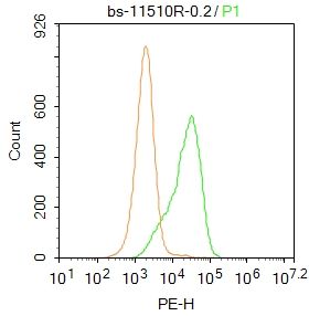
Blank control:A549.
Primary Antibody (green line): Rabbit Anti-TTC8 antibody (SL11510R)
Dilution: 1μg /10^6 cells;
Isotype Control Antibody (orange line): Rabbit IgG .
Secondary Antibody : Goat anti-rabbit IgG-PE
Dilution:0.2μg /test.
Protocol
The cells were fixed with 4% PFA (10min at room temperature)and then permeabilized with 20% PBST for 20 min at room temperature. The cells were then incubated in 5% BSA to block non-specific protein-protein interactions for 30 min at at room temperature .Cells stained with Primary Antibody for 30 min at room temperature. The secondary antibody used for 40 min at room temperature. Acquisition of 20,000 events was performed.
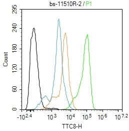
Blank control:SH-SY5Y.
Primary Antibody (green line): Rabbit Anti-Iba1 antibody (SL11510R)
Dilution: 2ug/Test;
Secondary Antibody : Goat anti-rabbit IgG-FITC
Dilution: 0.5ug/Test.
Protocol
The cells were fixed with 4% PFA (10min at room temperature)and then permeabilized with 90% ice-cold methanol for 20 min at -20℃.The cells were then incubated in 5%BSA to block non-specific protein-protein interactions for 30 min at room temperature .Cells stained with Primary Antibody for 30 min at room temperature. The secondary antibody used for 40 min at room temperature. Acquisition of 20,000 events was performed.
|
|
|