The gene encoding DYX1C1 maps in the 15q21 region, which is disrupted by a translocation t(2;15)(q11;q21) and segregates with dyslexia. Two sequence changes in DYX1C1, including one involving the translation initiation sequence and an Elk-1 transcription factor binding site (-3G --> A) and a codon (1249G --> T), introduce a premature stop codon and truncate the protein by 4 amino acids. DYX1C1 encodes a nuclear tetratricopeptide repeat domain protein that is dynamically regulated in brain. In human brain, DYX1C1 protein localizes to a fraction of cortical neurons and white matter glial cells. It is also expressed in lung, kidney and testis.
Function:
Involved in neuronal migration during development of the cerebral neocortex. May regulate the stability and proteasomal degradation of the estrogen receptors that play an important role in neuronal differentiation, survival and plasticity.
Subunit:
Interacts with ESR1 and ESR2. Interacts with STUB1.
Subcellular Location:
Nucleus. Cytoplasm.
Tissue Specificity:
Expressed in several tissues, including brain, lung, kidney and testis. In brain localizes to a fraction of cortical neurons and white matter glial cells.
DISEASE:
Defects in DYX1C1 may be a cause of susceptibility to dyslexia type 1 (DYX1) [MIM:127700]. A relatively common, complex cognitive disorder characterized by an impairment of reading performance despite adequate motivational, educational and intellectual opportunities. It is a multifactorial trait, with evidence for familial clustering and heritability. Note=A chromosomal aberration involving DYX1C1 has been found in a family affected by dyslexia. Translocation t(2;15)(q11;q21).
Similarity:
Contains 1 CS domain.
Contains 3 TPR repeats.
SWISS:
Q8WXU2
Gene ID:
161582
Database links:
Entrez Gene: 453458 Chimpanzee
Entrez Gene: 161582 Human
Omim: 608706 Human
SwissProt: Q863A7 Chimpanzee
SwissProt: Q863A5 Gorilla
SwissProt: Q8WXU2 Human
SwissProt: Q863A4 Orangutan
| Picture |
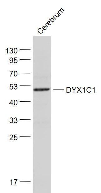
Sample:
Cerebrum (Mouse) Lysate at 40 ug
Primary: Anti- DYX1C1 (SL13043R) at 1/1000 dilution
Secondary: IRDye800CW Goat Anti-Rabbit IgG at 1/20000 dilution
Predicted band size: 49 kD
Observed band size: 49 kD
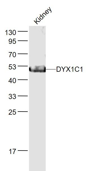
Sample:
Kidney (Mouse) Lysate at 40 ug
Primary: Anti- DYX1C1 (SL13043R) at 1/1000 dilution
Secondary: IRDye800CW Goat Anti-Rabbit IgG at 1/20000 dilution
Predicted band size: 49 kD
Observed band size: 49 kD
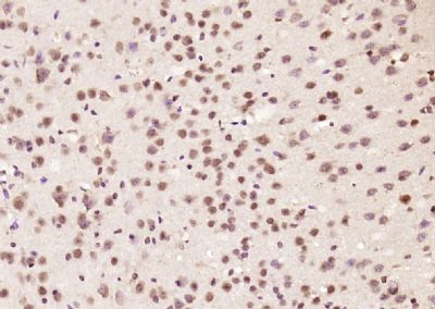
Paraformaldehyde-fixed, paraffin embedded (mouse brain); Antigen retrieval by boiling in sodium citrate buffer (pH6.0) for 15min; Block endogenous peroxidase by 3% hydrogen peroxide for 20 minutes; Blocking buffer (normal goat serum) at 37°C for 30min; Antibody incubation with (DYX1C1) Polyclonal Antibody, Unconjugated (SL13043R) at 1:200 overnight at 4°C, followed by operating according to SP Kit(Rabbit) (sp-0023) instructionsand DAB staining.
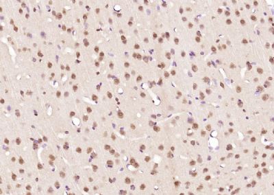
Paraformaldehyde-fixed, paraffin embedded (rat brain); Antigen retrieval by boiling in sodium citrate buffer (pH6.0) for 15min; Block endogenous peroxidase by 3% hydrogen peroxide for 20 minutes; Blocking buffer (normal goat serum) at 37°C for 30min; Antibody incubation with (DYX1C1) Polyclonal Antibody, Unconjugated (SL13043R) at 1:200 overnight at 4°C, followed by operating according to SP Kit(Rabbit) (sp-0023) instructionsand DAB staining.
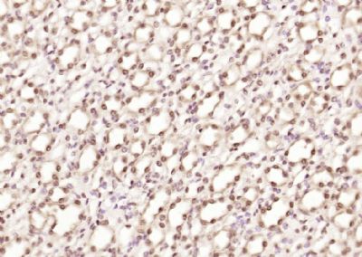
Paraformaldehyde-fixed, paraffin embedded (mouse kidney); Antigen retrieval by boiling in sodium citrate buffer (pH6.0) for 15min; Block endogenous peroxidase by 3% hydrogen peroxide for 20 minutes; Blocking buffer (normal goat serum) at 37°C for 30min; Antibody incubation with (DYX1C1) Polyclonal Antibody, Unconjugated (SL13043R) at 1:200 overnight at 4°C, followed by operating according to SP Kit(Rabbit) (sp-0023) instructionsand DAB staining.
|
|
|