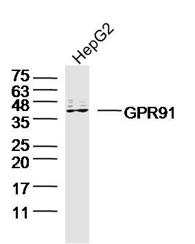
Sample: HepG2 Cell (Human) Lysate at 40 ug
Primary: Anti-GPR91(SL15392R)at 1/300 dilution
Secondary: IRDye800CW Goat Anti-Rabbit IgG at 1/20000 dilution
Predicted band size: 39kD
Observed band size: 39kD
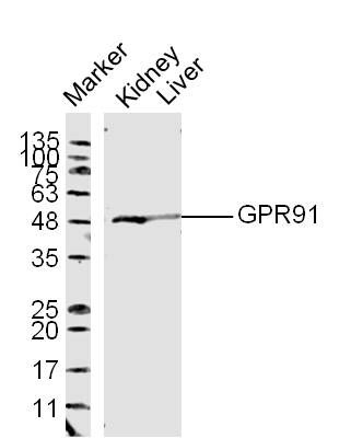
Protein: 1.kidney lyates(mo);2.liver lyates(mo);
Primary: Rabbit Anti-GPR91 (SL15392R) at 1:300;
Secondary: 800CW Conjugated Goat (polyclonal) Anti-Rabbit IgG(H+L) at 1: 10000;
Predicted band size:39 kD
Observed band size:49 kD
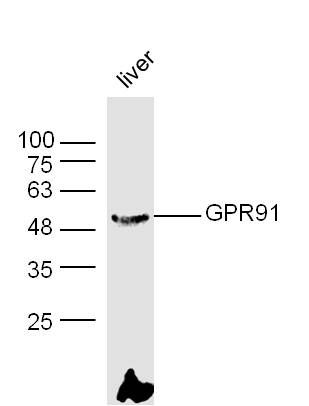
Western blot analysis of extracts from Liver tissue(mo) using GPR91 Antibody.
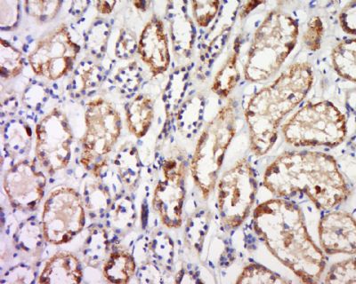
Tissue/cell: human kidney tissue; 4% Paraformaldehyde-fixed and paraffin-embedded;
Antigen retrieval: citrate buffer ( 0.01M, pH 6.0 ), Boiling bathing for 15min; Block endogenous peroxidase by 3% Hydrogen peroxide for 30min; Blocking buffer (normal goat serum,SLC0005) at 37℃ for 20 min;
Incubation: Anti-GPR91 Polyclonal Antibody, Unconjugated(SL15392R) 1:600, overnight at 4°C, followed by conjugation to the secondary antibody(SP-0023) and DAB(SLC0010) staining
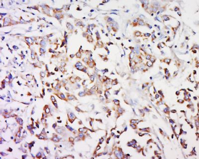
Tissue/cell: human breast cancer; 4% Paraformaldehyde-fixed and paraffin-embedded;
Antigen retrieval: citrate buffer ( 0.01M, pH 6.0 ), Boiling bathing for 15min; Block endogenous peroxidase by 3% Hydrogen peroxide for 30min; Blocking buffer (normal goat serum,SLC0005) at 37℃ for 20 min;
Incubation: Anti-GPR91 Polyclonal Antibody, Unconjugated(SL15392R) 1:600, overnight at 4°C, followed by conjugation to the secondary antibody(SP-0023) and DAB(SLC0010) staining
|