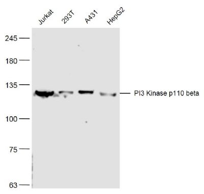
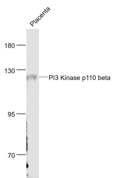
[IF=5.878] Lingling Dong. et al. Anti-inflammatory effect of Rhein on ulcerative colitis via inhibiting PI3K/Akt/mTOR signaling pathway and regulating gut microbiota. 2022 Mar 01 WB ; Mouse.
[IF=4.034] Siyuan Liu. et al. β-Hydroxybutyrate impairs the release of bovine neutrophil extracellular traps through inhibiting phosphoinositide 3-kinase–mediated nicotinamide adenine dinucleotide phosphate oxidase reactive oxygen species production. J Dairy Sci. 2022 Feb;: WB ; Cow.
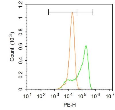
[IF=3.943] Le Tian. et al. Luteolin as an adjuvant effectively enhances CTL anti-tumor response in B16F10 mouse model. Int Immunopharmacol. 2021 May;94:107441 WB ; Mouse.
[IF=3.69] Yuling Tong. et al. Zuojin Pill ameliorates chronic atrophic gastritis induced by MNNG through TGF-β1/PI3K/Akt axis. J Ethnopharmacol. 2021 May;271:22893 WB ; Rat.
[IF=11.508] Qinyu Ma. et al. Osteoclast-derived apoptotic bodies couple bone resorption and formation in bone remodeling. Bone Res. 2021 Jan;9(1):1-12 WB ; Mouse.
[IF=2.563] Jiang S et al. Hesperetin as an adjuvant augments protective antitumor immunity responses in B16F10 melanoma by stimulating cytotoxic CD8+ T cells. Scandinavian Journal of Immunology, 2020: e12867. WB ; Mouse.
[IF=3.514] Li B et al. Resistin up-regulates LPL expression through the PPARγ-dependent PI3K/AKT signaling pathway impacting lipid accumulation in RAW264. 7 macrophages.Cytokine. 2019 Jul;119:168-174. WB ; Mouse.
[IF=1.41] Wei R et al. Astragaloside Ⅳ combating liver cirrhosis through the PI3K/Akt/mTOR signaling pathway. EXPERIMENTAL AND THERAPEUTIC MEDICINE.2019 17: 393-397. WB ; Rat.
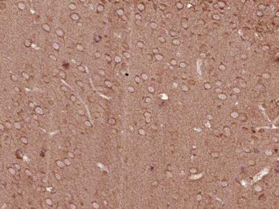
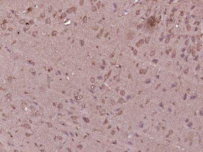
[IF=4.292] Wenqian Xie. et al. Seasonal expressions of ERα, ERβ, EGF, EGFR, PI3K and Akt in the scent glands of the muskrats (Ondatra zibethicus). J Steroid Biochem. 2021 Oct;213:105961 IHC ; Muskrat.
[IF=6.304] Xuemei Shen. et al. CircRILPL1 promotes muscle proliferation and differentiation via binding miR-145 to activate IGF1R/PI3K/AKT pathway. Cell Death Dis. 2021 Feb;12(2):1-14 WB ; Bovine.