Home > Product > Antibody > Rabbit Anti-MC1R antibody
MC1 Receptor; CMM5; MC1-R; MC-1R; Melanocortin 1 receptor; Melanocortin 1 receptor (alpha melanocyte stimulating hormone receptor); Melanocortin receptor 1; Melanocyte-stimulating hormone receptor; Melanotropin receptor; MSH-R; MSHR; MSHR_HUMAN; SHEP2.
Cat:
SL23517R
Species Reactivity:
Human,
Immunogen:
KLH conjugated synthetic peptide derived from human MC1 Receptor :181-280/317<Cytoplasmic>
Format:
Liquid
Storage instructions:
Shipped at 4℃. Store at -20 °C for one year. Avoid repeated freeze/thaw cycles.
Concentration:
1mg/ml
Clonality:
Polyclonal
Isotype:
IgG
Applications:
WB=1:500-2000ELISA=1:5000-10000IHC-P=1:100-500IHC-F=1:100-500ICC=1:100-500IF=1:100-500(Paraffin sections need to do antigen repair)not yet tested in other applications.optimal dilutions/concentrations should be determined by the end user.
Host:
Rabbit
Product Overview:
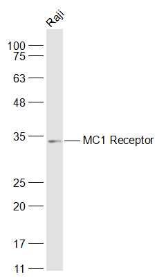
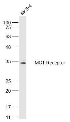
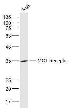
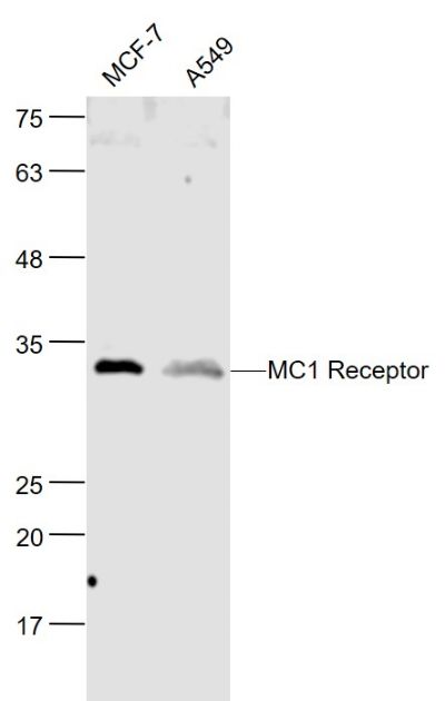
Sample: Raji(Human) Cell Lysate at 30 ugPrimary: Anti-MC1 Receptor (SL23517R) at 1/1000 dilutionSecondary: IRDye800CW Goat Anti-Rabbit IgG at 1/20000 dilutionPredicted band size: 35 kDObserved band size: 34 kDSample: MOLT-4(Human) Cell Lysate at 30 ugPrimary: Anti-MC1 Receptor (SL23517R) at 1/1000 dilutionSecondary: IRDye800CW Goat Anti-Rabbit IgG at 1/20000 dilutionPredicted band size: 35 kDObserved band size: 34 kDSample: Raji(Human) Cell Lysate at 30 ugPrimary: Anti-MC1 Receptor (SL23517R) at 1/1000 dilutionSecondary: IRDye800CW Goat Anti-Rabbit IgG at 1/20000 dilutionPredicted band size: 35 kDObserved band size: 35 kDSample: MCF-7 (Human) Cell Lysate at 30 ugA549 (Human) Cell Lysate at 30 ugPrimary: Anti- MC1 Receptor (SL23517R) at 1/1000 dilutionSecondary: IRDye800CW Goat Anti-Rabbit IgG at 1/20000 dilutionPredicted band size: 35 kDObserved band size: 34 kDParaformaldehyde-fixed, paraffin embedded (Human melanoma); Antigen retrieval by boiling in sodium citrate buffer (pH6.0) for 15min; Block endogenous peroxidase by 3% hydrogen peroxide for 20 minutes; Blocking buffer (normal goat serum) at 37°C for 30min; Antibody incubation with (MC1) Polyclonal Antibody, Unconjugated (SL23517R) at 1:400 overnight at 4°C, followed by operating according to SP Kit(Rabbit) (sp-0023) instructionsand DAB staining.
More
Unit:
Price: $
Product PDFs
Datasheet:


This intronless gene encodes the receptor protein for melanocyte -stimulating hormone (MSH). The encoded protein, a seven pass transmembrane G protein coupled receptor, controls melanogenesis. Two types of melanin exist: red pheomelanin and black eumelanin. Gene mutations that lead to a loss in function are associated with increased pheomelanin production, which leads to lighter skin and hair color. Eumelanin is photoprotective but pheomelanin may contribute to USLVinduced skin damage by generating free radicals upon UV radiation. Binding of MSH to its receptor activates the receptor and stimulates eumelanin synthesis. This receptor is a major determining factor in sun sensitivity and is a genetic risk factor for melanoma and non-melanoma skin cancer. Over30 variant alleles have been identified which correlate with skin and hair color, providing evidence that this gene is an important component in determining normal human pigment variation.

Function:
Receptor for MSH (alpha, beta and gamma) and ACTH. The activity of this receptor is mediated by G proteins which activate adenylate cyclase.

Subcellular Location:
Cell membrane.

Tissue Specificity:
Melanocytes and corticoadrenal tissue.

DISEASE:
Genetic variations in MC1R are a cause of susceptibility to cutaneous malignant melanoma type 5 (CMM5) [MIM:613099]. Malignant melanoma is a malignant neoplasm of melanocytes, arising de novo or from a pre-existing benign nevus, which occurs most often in the skin but also may involve other sites.

Similarity:
Belongs to the G-protein coupled receptor 1 family.

SWISS:
Q01727

Gene ID:
4157

Database links:

Entrez Gene: 4157 Human

Omim: 155555 Human

SwissProt: Q01726 Human

Unigene: 513829 Human


类固醇受体(Steroid Receptors) Picture

Sample:
Raji(Human) Cell Lysate at 30 ug
Primary: Anti-MC1 Receptor (SL23517R) at 1/1000 dilution
Secondary: IRDye800CW Goat Anti-Rabbit IgG at 1/20000 dilution
Predicted band size: 35 kD
Observed band size: 34 kD
Sample:
MOLT-4(Human) Cell Lysate at 30 ug
Primary: Anti-MC1 Receptor (SL23517R) at 1/1000 dilution
Secondary: IRDye800CW Goat Anti-Rabbit IgG at 1/20000 dilution
Predicted band size: 35 kD
Observed band size: 34 kD
Sample:
Raji(Human) Cell Lysate at 30 ug
Primary: Anti-MC1 Receptor (SL23517R) at 1/1000 dilution
Secondary: IRDye800CW Goat Anti-Rabbit IgG at 1/20000 dilution
Predicted band size: 35 kD
Observed band size: 35 kD
Sample:
MCF-7 (Human) Cell Lysate at 30 ug
A549 (Human) Cell Lysate at 30 ug
Primary: Anti- MC1 Receptor (SL23517R) at 1/1000 dilution
Secondary: IRDye800CW Goat Anti-Rabbit IgG at 1/20000 dilution
Predicted band size: 35 kD
Observed band size: 34 kD
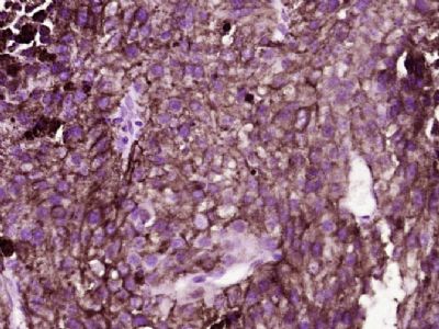
Paraformaldehyde-fixed, paraffin embedded (Human melanoma); Antigen retrieval by boiling in sodium citrate buffer (pH6.0) for 15min; Block endogenous peroxidase by 3% hydrogen peroxide for 20 minutes; Blocking buffer (normal goat serum) at 37°C for 30min; Antibody incubation with (MC1) Polyclonal Antibody, Unconjugated (SL23517R) at 1:400 overnight at 4°C, followed by operating according to SP Kit(Rabbit) (sp-0023) instructionsand DAB staining.
Product Feedback Wall
Message :
Your Email :
Copyright © 2007-2018 Sunlong Medical All Rights Reserved.